

Deep learning from "passive feeding" to "selective eating" of real-world data
- PMID: 33145439
- PMCID: PMC7603327
- DOI: 10.1038/s41746-020-00350-y
Deep learning from "passive feeding" to "selective eating" of real-world data
Abstract
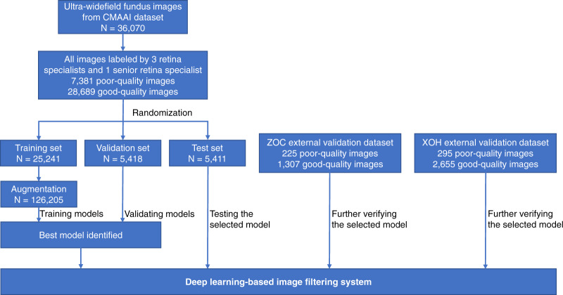
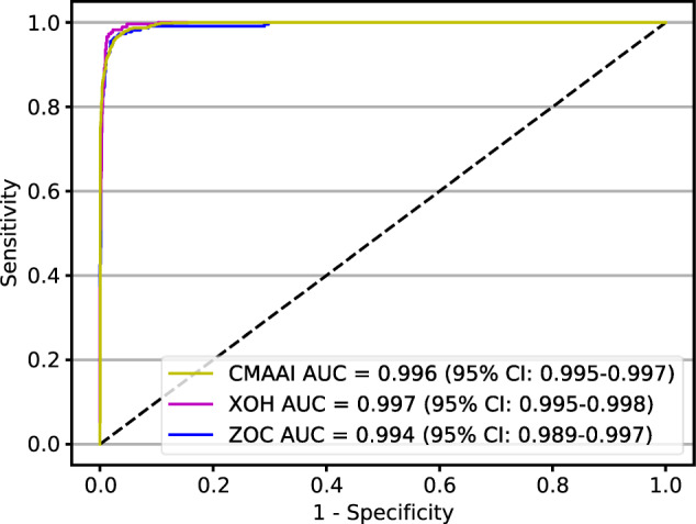
Artificial intelligence (AI) based on deep learning has shown excellent diagnostic performance in detecting various diseases with good-quality clinical images. Recently, AI diagnostic systems developed from ultra-widefield fundus (UWF) images have become popular standard-of-care tools in screening for ocular fundus diseases. However, in real-world settings, these systems must base their diagnoses on images with uncontrolled quality ("passive feeding"), leading to uncertainty about their performance. Here, using 40,562 UWF images, we develop a deep learning-based image filtering system (DLIFS) for detecting and filtering out poor-quality images in an automated fashion such that only good-quality images are transferred to the subsequent AI diagnostic system ("selective eating"). In three independent datasets from different clinical institutions, the DLIFS performed well with sensitivities of 96.9%, 95.6% and 96.6%, and specificities of 96.6%, 97.9% and 98.8%, respectively. Furthermore, we show that the application of our DLIFS significantly improves the performance of established AI diagnostic systems in real-world settings. Our work demonstrates that "selective eating" of real-world data is necessary and needs to be considered in the development of image-based AI systems.
Keywords: Medical imaging; Translational research.
© The Author(s) 2020.
Conflict of interest statement
Competing interestsThe authors declare no competing interests.
Figures


Similar articles
-
Development of a deep learning-based image eligibility verification system for detecting and filtering out ineligible fundus images: A multicentre study.Int J Med Inform. 2021 Mar;147:104363. doi: 10.1016/j.ijmedinf.2020.104363. Epub 2020 Dec 13. Int J Med Inform. 2021. PMID: 33388480
-
Deep learning for automated glaucomatous optic neuropathy detection from ultra-widefield fundus images.Br J Ophthalmol. 2021 Nov;105(11):1548-1554. doi: 10.1136/bjophthalmol-2020-317327. Epub 2020 Sep 16. Br J Ophthalmol. 2021. PMID: 32938630
-
Development of a deep learning-based image quality control system to detect and filter out ineligible slit-lamp images: A multicenter study.Comput Methods Programs Biomed. 2021 May;203:106048. doi: 10.1016/j.cmpb.2021.106048. Epub 2021 Mar 17. Comput Methods Programs Biomed. 2021. PMID: 33765481
-
Ultra-Widefield Imaging for Evaluation of the Myopic Eye.Semin Ophthalmol. 2021 May 19;36(4):185-190. doi: 10.1080/08820538.2021.1887904. Epub 2021 Feb 23. Semin Ophthalmol. 2021. PMID: 33620294 Review.
-
AI applications to medical images: From machine learning to deep learning.Phys Med. 2021 Mar;83:9-24. doi: 10.1016/j.ejmp.2021.02.006. Epub 2021 Mar 1. Phys Med. 2021. PMID: 33662856 Review.
Cited by
-
Comparison of deep learning systems and cornea specialists in detecting corneal diseases from low-quality images.iScience. 2021 Oct 22;24(11):103317. doi: 10.1016/j.isci.2021.103317. eCollection 2021 Nov 19. iScience. 2021. PMID: 34778732 Free PMC article.
-
Preventing corneal blindness caused by keratitis using artificial intelligence.Nat Commun. 2021 Jun 18;12(1):3738. doi: 10.1038/s41467-021-24116-6. Nat Commun. 2021. PMID: 34145294 Free PMC article.
-
Digital ray: enhancing cataractous fundus images using style transfer generative adversarial networks to improve retinopathy detection.Br J Ophthalmol. 2024 Sep 20;108(10):1423-1429. doi: 10.1136/bjo-2024-325403. Br J Ophthalmol. 2024. PMID: 38839251 Free PMC article. Clinical Trial.
-
Synergizing Deep Learning-Enabled Preprocessing and Human-AI Integration for Efficient Automatic Ground Truth Generation.Bioengineering (Basel). 2024 Apr 28;11(5):434. doi: 10.3390/bioengineering11050434. Bioengineering (Basel). 2024. PMID: 38790302 Free PMC article.
-
Quickly diagnosing Bietti crystalline dystrophy with deep learning.iScience. 2024 Jul 25;27(9):110579. doi: 10.1016/j.isci.2024.110579. eCollection 2024 Sep 20. iScience. 2024. PMID: 39220263 Free PMC article.
References
LinkOut - more resources
Full Text Sources
