

Accessory Nerve Anatomy in Anterior and Posterior Cervical Triangle: A Fresh Cadaveric Study
- PMID: 33145498
- PMCID: PMC7580510
- DOI: 10.5152/tao.2020.5263
Accessory Nerve Anatomy in Anterior and Posterior Cervical Triangle: A Fresh Cadaveric Study
Abstract
Objective: To understand the variations and normal course of the accessory nerve (CNXI) to help more accurate and confident neck dissection.
Methods: The course of the CNXI in the neck, its relationship to the surrounding anatomic structures and the factors affecting its course were investigated.
Results: A total of 100 neck dissections were performed on 50 fresh cadavers. Eleven division variations were observed at the anterior triangle. The location of CNXI at the posterior border of the sternocleidomastoid muscle (PBSCM) was investigated and the ratio between the distance from the mastoid apex (MAA) to CNXI at the PBSCM and the distance from MAA to the posterior border where the PBSCM is attached to the clavicle increased as height of the subject increased (p<0.05).
Conclusion: It must be kept in mind that it is better to search for CNXI in taller subjects more inferiorly at the posterior border of the sternocleidomastoid muscle.
Keywords: Accessory nerve; Erb’s point; anatomy; injury; neck dissection; sternocleidomastoid muscle.
© Copyright 2020 by Official Journal of the Turkish Society of Otorhinolaryngology and Head and Neck Surgery.
Conflict of interest statement
Conflict of Interest: The authors have no conflicts of interest to declare.
Figures
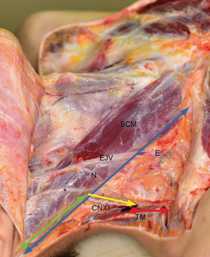
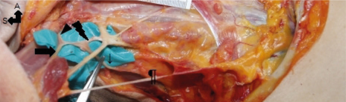
 : accessory nerve; thin arrow: trapezial branch of accessory nerve; thick arrow: sternocleidomastoid branch of accessory nerve; lightning: connection between CNXI and C2
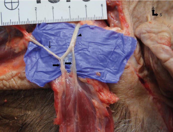 : accessory nerve; thin arrow: trapezial branch of accessory nerve; thick arrow: sternocleidomastoid branch of accessory nerve
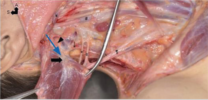 : accessory nerve; thin arrow: trapezial branch of accessory nerve; thick arrow: sternocleidomastoid branch of accessory nerve; *: great auricular nerve; ǂ: cutaneous branches of the cervical plexus nerves; blue arrow: PBD-SCMen
Similar articles
-
Spinal accessory nerve anatomy in the posterior cervical triangle: A systematic review with meta-analysis.Clin Anat. 2024 Jan;37(1):130-139. doi: 10.1002/ca.24119. Epub 2023 Sep 28. Clin Anat. 2024. PMID: 37767816
-
Anatomical study of phrenic nerve course in relation to neck dissection.Surg Radiol Anat. 2015 Apr;37(3):255-8. doi: 10.1007/s00276-014-1343-1. Epub 2014 Jul 16. Surg Radiol Anat. 2015. PMID: 25026999
-
Vulnerability of the spinal accessory nerve in the posterior triangle of the neck: a cadaveric study.Orthopedics. 2002 Jan;25(1):71-4. doi: 10.3928/0147-7447-20020101-20. Orthopedics. 2002. PMID: 11811246
-
Spinal accessory nerve preservation in modified neck dissections: surgical and functional outcomes.Acta Otorhinolaryngol Ital. 2017 Oct;37(5):368-374. doi: 10.14639/0392-100X-844. Acta Otorhinolaryngol Ital. 2017. PMID: 29165431 Free PMC article.
-
Anterior approach to the cervical spine: surgical anatomy.Orthopedics. 2000 Aug;23(8):841-5. doi: 10.3928/0147-7447-20000801-19. Orthopedics. 2000. PMID: 10952048 Review.
Cited by
-
Anatomic Variability of the Accessory Nerve: Implications for Dissection of Level IIB.Laryngoscope. 2024 Jan;134(1):154-159. doi: 10.1002/lary.30758. Epub 2023 Jun 8. Laryngoscope. 2024. PMID: 37289066 Free PMC article.
-
Super-Superselective Level VB Neck Dissection for Papillary Thyroid Cancer.Cancers (Basel). 2025 Apr 29;17(9):1497. doi: 10.3390/cancers17091497. Cancers (Basel). 2025. PMID: 40361424 Free PMC article. Review.
-
Study of the Anatomical Variations of Spinal Accessory Nerve Seen During Neck Dissection.Indian J Otolaryngol Head Neck Surg. 2024 Jun;76(3):2295-2303. doi: 10.1007/s12070-023-04468-9. Epub 2024 Jan 18. Indian J Otolaryngol Head Neck Surg. 2024. PMID: 38883541 Free PMC article.
References
-
- Dargent M, Papillon J. Motor complications of neck dissection: how to avoid them. Lyon Chir. 1945;40:718. - PubMed
LinkOut - more resources
Full Text Sources
Miscellaneous