. 2021 Apr;238(4):1010-1022.
doi: 10.1111/joa.13344. Epub 2020 Nov 3.

Initial morphological symmetry breaking in the foregut and development of the omental bursa in human embryos

Affiliations

Initial morphological symmetry breaking in the foregut and development of the omental bursa in human embryos

Tobias Schäfer et al. J Anat. 2021 Apr.

Abstract

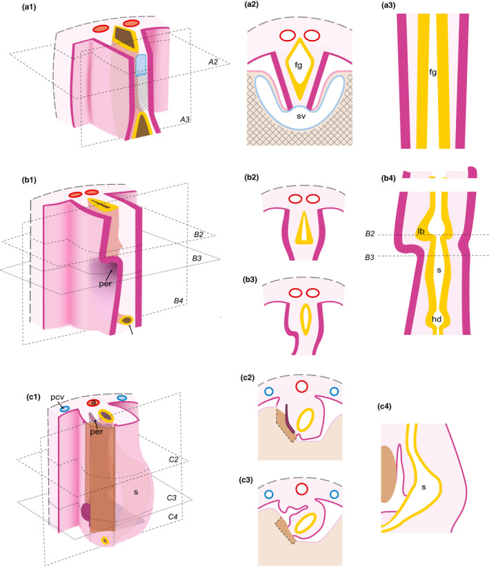
Bilaterally symmetrical primordia of visceral organs undergo asymmetrical morphogenesis leading to typical arrangement of visceral organs in the adult. Asymmetrical morphogenesis within the upper abdomen leads, among others, to the formation of the omental bursa dorsally to the rotated stomach. A widespread view of this process assumes kinking of thin mesenteries as a main mechanism. This view is based on a theory proposed already by Johannes Müller in 1830 and was repeatedly criticized, but some of the most plausible alternative views (initially proposed by Swaen in 1897 and Broman in 1904) still remain to be proven. Here, we analyzed serial histological sections of human embryos between stages 12 and 15 at high light microscopical resolution to reveal the succession of events giving rise to the development of the omental bursa and its relation to the emerging stomach asymmetry. Our analysis indicates that morphological symmetry breaking in the upper abdomen occurs within a wide mesenchymal plate called here mesenteric septum and is based on differential behavior of the coelomic epithelium which causes asymmetric paragastric recess formation and, importantly, precedes initial rotation of stomach. Our results thus provide the first histological evidence of breaking the symmetry of the early foregut anlage in the human embryo and pave the way for experimental studies of left-right symmetry breaking in the upper abdomen in experimental model organisms.

Keywords: coelom; left-right symmetry breaking; mesentery; omental bursa; stomach.


Figures

Figure 1
Figure 1
The mesenteric septum of an early stage 12 human embryo (Carnegie #8943). (a–d) Transversal sections. Intersecting arrows in A indicate anatomical axes. Asterisks mark the thickened coelomic epithelium. An overview image of the embryo with black lines indicating the position of the transversal sections is shown in the upper right corner of (d). (e, f) Magnification of the mesenteric septum and the developing foregut. Squared segments are shown at higher magnification in (b) and (c), respectively. Arrowheads indicate mitotic cells in the coelomic epithelium. White arrow points to the irregularly shaped basal side of the epithelium in (e) and black arrow points to the regular continuous line corresponding to the intact basal membrane in (f). Abbreviations: d, dorsal; v, ventral; l, left; r, right; ccc, common coelomic cavity (corresponds to intra‐embryonic coelom with exception of cavities formed by recesses); ao, aorta; sv, sinus venosus; st, septum transversum; ms, mesenteric septum; fg, foregut; mg, midgut; hd, hepatic diverticulum; and gb, gallbladder. Scale bar in (d) 100 µm for (a–d) and 20 µm for (e) and (f)
Figure 2
Figure 2
Asymmetric bud‐like thickening in the coelomic epithelium of the mesenteric septum as a first sign of symmetry breaking of a stage‐12 human embryo (Carnegie #8505A). (a–d) Transversal sections of the embryo (anatomical axes oriented as in Figure 1). the position of the transversal sections is indicated in the overview image in (d). Bars in (c) mark the position of the frontal sections shown in Figure 7(a) and (b). Abbreviations: ccc, common coelomic cavity; lb, lung bud; rv, right ventricle; lv, left ventricle; sv, sinus venosus; st, septum transversum; ms, mesenteric septum; and hd, hepatic diverticulum. Scale bar in (d): 100 µm for (a–d)
Figure 3
Figure 3
Early development of the pneumato‐enteric recess in an early‐stage‐13 embryo (#EB 1954_09_11). (a–c) Fronto‐transversal sections. Arrows point to the emerging right‐sided recess. The position of the fronto‐transversal sections is indicated in the overview image in (c). (d) High magnification of the squared segment in (c). Note the wide intercellular spaces within the mesenteric septum. Asterisk points to intercellular spaces formed by mesenchymal cells. Hash marks a blood vessel. Abbreviations: ao, aorta; ms, mesenteric septum; lb, lung bud; fg, foregut; and ccc, common coelomic cavity. Scale bar in (d): 100 µm for (a–c); 20 µm for (d)
Figure 4
Figure 4
Fully developed pneumato‐enteric recess in a stage‐13 embryo (Carnegie #836) next to an almost symmetrical stomach anlage. (a–e) Transversal sections of the embryo (anatomical axes are oriented as in Figure 1). Arrowhead in (c) points to the narrow invagination of the coelomic epithelium on the left side (cf. Figure 7). Position of the transversal sections is indicated in the overview image in (e). Abbreviations: ao, aorta; lb, lung bud; ms, mesenteric septum; pm, paramesentery; per, pneumato‐enteric recess; ra, right atrium; la, left atrium; rv, right ventricle; lv, left ventricle; sv, sinus venosus; st, septum transversum; and hc, hepatic cords. Scale bar in (e): 200 µm for (a–e)
Figure 5
Figure 5
Different epithelial configurations in the lining of the pneumato‐enteric recess shown in Figure 4 as signs of local morphogenetic epithelial activity. Sections are aligned according to the orientation of the longest cross‐sectional diameter of the recess. Intersecting arrows in A indicate anatomical axes. Arrows point to the endothelial covering of blood vessels; and hashes mark the cleft which corresponds to the lumen of the blood vessel. Arrowhead in (b) shows a mitotic figure. Black square brackets mark the height of columnar (pseudostratified) and flat stretches of the epithelium, respectively. Scale bar in (d): 20 µm for (a–c)
Figure 6
Figure 6
Increasing stomach asymmetry correlates with the expansion of the mesenterico‐enteric recess. (a–d) Transversal sections of a stage‐15 embryo (embryo Carnegie #3512). Anatomical axes are oriented as in Figure 1. Asterisk marks the lumen connecting the common coelomic cavity with the developing omental bursa. Position of the transversal sections is indicated in the overview image in (d). Abbreviations: ccc, common coelomic cavity; a, aorta; lb, lung bud; e, esophagus; per, her, and mer/per, (right) pneumato‐enteric, hepato‐enteric, and mesenterico‐enteric (pancreatico‐enteric) recesses, respectively; s, stomach; d, duodenum; dm, dorsal mesogastrium; hc, hepatic cords; and pm, paramesentery. Scale bar in (d): 50 µm for (a–d)
Figure 7
Figure 7
Foregut asymmetry between stages 12 (a–c) and 13 (d–f): The asymmetrically arranged mesenteric septum precedes the emergence of the pneumato‐enteric recess. (a, b) Frontal views of 3D reconstructions of a stage‐12 embryo (Carnegie #8505A); (b) is, in addition, rotated counter‐clockwise in the transversal plane to show the invagination in the coelomic epithelium (arrow). (c) Right lateral view of the visceral coelomic epithelial covering at stage 12. (d) Frontal view of 3D reconstruction of the coelomic cavity and the pneumato‐enteric recess at stage 13 (embryo Carnegie #836). Arrowhead in (c) corresponds to the narrow invagination on the left side seen in Figure 4. (e) Asymmetrical arrangement of coelomic epithelium and coelomic cavities seen in the frontal section of 3D reconstruction of the same stage 13 embryo. Formation of paramesenteric fold (f) on the right side (cf. Figure S1). (f) Right lateral view of the visceral coelomic epithelial covering at stage 13 demonstrating the narrow opening of the emerging pneumato‐enteric recess (white arrow). Stomach (s) and lung bud (lb) are in green; coelomic epithelium (ce) in azure blue; common coelomic cavity (ccc) in turquoise; pneumato‐enteric recess (per) in teal; surface of section through epithelium in gray, and through common coelomic cavity (ccc), in turquoise
Figure 8
Figure 8
Ventral and dorsal views of isolated stomach and omental bursa anlagen, the latter of which is composed of the pneumato‐enteric (per), the mesenterico‐enteric (mer), and the hepato‐enteric (her) recesses, at stage 15 and derived from the 3D reconstructed embryo shown in Figure 6 (Carnegie #3512). The omental bursa is located right and dorso caudal to the stomach which displays significant left‐right asymmetry with major and minor curvatures on its left and right sides, respectively. Stomach (s) and esophagus (e) are presented in green, omental bursa primordia in teal
Figure 9
Figure 9
Steps of omental bursa development inferred from the results of the present study. Related areas are shown in anterolateral views (a1; b1; c1) and transversal (a2; b2; b3; c2; c3) and frontal (a3; b4; c4) sections. (a1‐a3) Initial symmetrical arrangement of the foregut and its mesenteries. (b1‐b4) Beginning of left‐right asymmetry in the mesenteric septum. (c1‐c4) Asymmetric enlargement of recesses and corresponding displacement of the stomach primordium. Structures located ventrally (including especially septum transversum and liver primordia as well as the sinus venosus) are removed in a1 to enable the view into the coelomic cavity. Abbreviations: s, stomach; lb, lung bud; hd, hepatic diverticulum; a, aorta; and sv, sinus venosus. Crosshatched lines illustrate septum transversum. Epithelial covering of the coelom, pink; sectioned coelomic epithelium, magenta; mesenchymal tissue of the digestive system and the neighboring posterior body wall, rose; aorta, red; veins, blue; foregut with lung and hepatic buds, yellow; and liver primordium tissue, brown. The levels of transversal and frontal sections are shown in a1, b1, and c1, respectively

References

    1. Aben, K. & de Bakker, B.S. (2012) The embryonic development of the pneumato‐enteric recess. Amsterdam: Amsterdam University College.
    1. Bartram, U. , Wirbelauer, J. & Speer, C.P. (2005) Heterotaxy syndrome – Asplenia and polysplenia as indicators of visceral malposition and complex congenital heart disease. Biology of the Neonate, 88, 278–290. - PubMed
    1. Blum, M. & Ott, T. (2018) Animal left‐right asymmetry. Current Biology, 28, R301–R304. - PubMed
    1. Broman, I. (1904) Die Entwicklungsgeschichte der Bursa omentalis und aehnlicher Rezessbildungen bei den Wirbeltieren. Wiesbaden: Bergmann.
    1. Broman, I. (1905) Über die Entwickelung der Mesenterien der Leberligamente und der Leberform bei den Lungenfischen. Jena, Germany: Fischer.

Publication types