. 2022 May;12(5):1050-1060.
doi: 10.1002/2211-5463.13022. Epub 2022 Mar 29.

Prefoldin subunit 6 of Plasmodium falciparum binds merozoite surface protein-1

Affiliations

Prefoldin subunit 6 of Plasmodium falciparum binds merozoite surface protein-1

Vikash Kumar et al. FEBS Open Bio. 2022 May.

Abstract

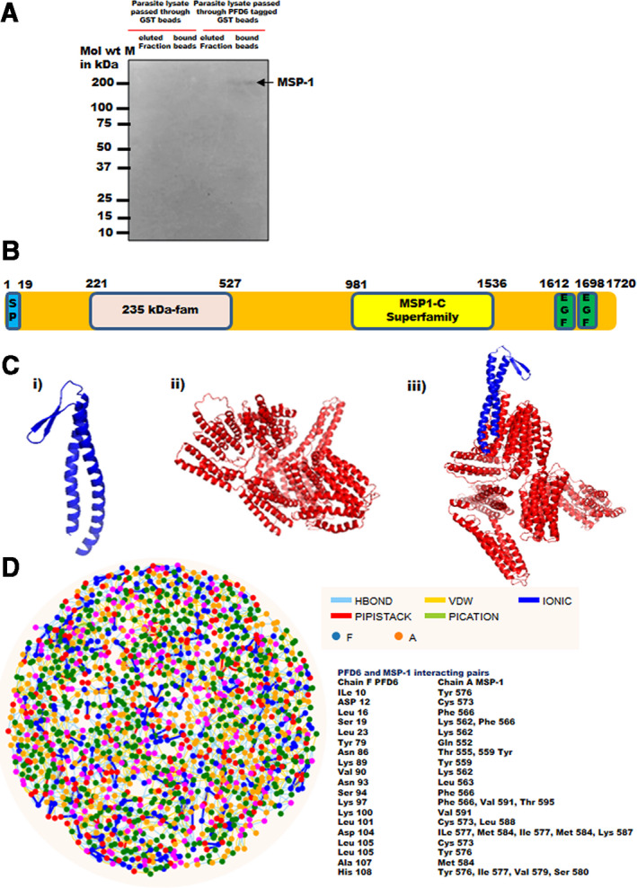
Malaria is a human disease caused by eukaryotic protozoan parasites of the Plasmodium genus. Plasmodium falciparum (Pf) causes the most lethal form of human malaria and is responsible for widespread mortality worldwide. Prefoldin is a heterohexameric molecular complex that binds and delivers unfolded proteins to chaperonin for correct folding. The prefoldin PFD6 is predicted to interact with merozoite surface protein-1 (MSP-1), a protein well known to play a pivotal role in erythrocyte binding and invasion by Plasmodium merozoites. We previously found that the P. falciparum (Pf) genome contains six prefoldin genes and a prefoldin-like gene whose molecular functions are unidentified. Here, we analyzed the expression of PfPFD-6 during the asexual blood stages of the parasite and investigated its interacting partners. PfPFD-6 was found to be significantly expressed at the trophozoite and schizont stages. Pull-down assays suggest PfPFD-6 interacts with MSP-1. In silico analysis suggested critical residues involved in the PfPFD-6-MSP-1 interaction. Our data suggest PfPFD-6 may play a role in stabilizing or trafficking MSP-1.

Keywords: Plasmodium falciparum; chaperone; malaria; merozoite surface protein-1; prefoldin.


Conflict of interest statement

The authors declare no conflict of interest.

Figures

Fig. 1
Fig. 1
Domain organization and expression of PfPFD‐6 protein. (A) Domain organization of PF3D7_0512000 (PfPFD‐6). Schematic representation of full‐length PfPFD‐6 protein (yellow) containing a prefoldin β‐superfamily domain (blue) (B) SDS/PAGE of purified recombinant protein cloned in pET28a vector lane 1: protein ladder; lane 2: purified recombinant PfPFD‐6. Dashed line indicates that lanes were not originally adjacent. (C) SDS/PAGE of purified recombinant protein cloned in pGEX4T‐1 vector. Lane 1: protein ladder; lane 2: purified recombinant PfPFD‐6 (D) Western blot of purified recombinant protein using antihistidine antibodies. (E) Western blot of purified recombinant protein using anti‐GST antibodies.
Fig. 2
Fig. 2
Multiple sequence alignment and phylogenetic tree of PfPFD‐6 subunits with its homologs. (A) Comparison of amino acid sequence of PfPFD‐6 subunits with its homologs in Plasmodium species, Homo sapiens, T. gondii, C. elegans, Saccharomyces cerevisiae, A. thaliana, M. musculus, and C. lupus. Alignment was performed using clustal omega, EMBL‐EBI, Hinxton, Cambridge, UK. ‘*’ represents conserved residues, whereas ‘:’ and ‘.’ represent semi‐conserved residues. (B) Phylogenetic tree of PfPFD‐6 subunits with its homologs using mega 6 software, Pennsylvania State University, PA, USA.
Fig. 3
Fig. 3
In vivo expression and colocalization assay. (A) Western blot analysis of schizont stage Pf3D7 parasite lysates to test in vivo expression of PfPFD‐6. Lane 1: protein ladder; lane 2: schizont stage parasite lysate. Blot was probed with anti‐PfPFD‐6 antibodies followed by secondary antibodies. (B) Expression analysis and colocalization of PfPFD‐6 with MSP‐1 at different asexual stages of parasite life cycle. Smears of methanol‐fixed Pf3D7‐infected erythrocytes were stained with anti‐PfPFD‐6 antibodies (1 : 250) and anti‐MSP‐1 antibodies (1 : 250), followed by incubation with Alexa Fluor‐conjugated secondary antibodies (Alexa Fluor 488, red; Alexa Fluor 546, green). DIC, differential interference contrast image; DAPI, nuclear staining 40, 6‐diamidino‐2‐phenylindole (blue); MSP‐1, mouse anti‐MSP‐1 (green); PfPFD‐6, anti‐PfPFD‐6 antibody (red); merge, overlay of PfPFD‐6 proteins with MSP‐1. Scale bar represents 2 μm.
Fig. 4
Fig. 4
Domain organization of MSP‐1 protein and interaction of PfPFD‐6 with MSP‐1. (A) Pull‐down assay of PfPFD‐6 with MSP‐1. Beads bound with GST‐tagged PfPFD‐6 and only GST‐bound beads were incubated with schizont stage parasite lysate. The protein complex was recovered from the beads by eluting in elution buffer. Eluted fraction and protein complex bound beads were resolved on 12% SDS/PAGE, transferred to nitrocellulose membrane, and probed with monoclonal anti‐MSP119 (1 : 500). Lane M: protein ladder, lane 1: eluted fraction of GST‐bound beads, lane 2: supernatant of boiled GST‐bound beads, lane 3: eluted fraction of GST‐tagged PfPFD‐6‐bound beads, and lane 4: supernatant of boiled GST‐tagged PfPFD‐6‐bound beads. (B) Schematic representation of MSP‐1 showing signal peptide (SP, blue), 235 kDa fam domain (pink), MSP1‐C superfamily domain (yellow), and EGF domain (green). (C) Cartoon representation of modeled structures. (i) Model structure of PfPFD‐6, (ii) model structure of MSP‐1, and (iii) docked structure of PfPFD‐6‐MSP‐1 complex. (D) RIN plot of PfPFD‐6‐MSP‐docked complex. Nodes in blue and orange on the plot represent residues of PfPFD‐6 and MSP‐1, respectively. Pairs of interacting residues are mentioned on the plot (right panel).

Similar articles

Cited by

References

    1. Gardner MJ, Hall N, Fung E, White O, Berriman M, Hyman RW, Carlton JM, Pain A, Nelson KE, Bowman S et al. (2002) Genome sequence of the human malaria parasite Plasmodium falciparum . Nature 419, 498–511. - PMC - PubMed
    1. Cox FE (2010) History of the discovery of the malaria parasites and their vectors. Parasites & Vectors 3, 1–9. - PMC - PubMed
    1. Sinnis P and Zavala F (2012) The skin: where malaria infection and the host immune response begin. Semin Immunopathol 34, 787–792. - PMC - PubMed
    1. Hafalla JC, Silvie O and Matuschewski K (2011) Cell biology and immunology of malaria. Immunol Rev 240, 297–316. - PubMed
    1. Perkins ME and Rocco LJ (1988) Sialic acid‐dependent binding of Plasmodium falciparum merozoite surface antigen, Pf200, to human erythrocytes. J Immunol 141, 3190–3196. - PubMed