Review
. 2020 Nov 13;134(21):2851-2871.
doi: 10.1042/CS20200899.

ACE2 and ACE: structure-based insights into mechanism, regulation and receptor recognition by SARS-CoV

Affiliations
Review

ACE2 and ACE: structure-based insights into mechanism, regulation and receptor recognition by SARS-CoV

Lizelle Lubbe et al. Clin Sci (Lond). .

Abstract

Angiotensin converting enzyme (ACE) is well-known for its role in blood pressure regulation via the renin-angiotensin aldosterone system (RAAS) but also functions in fertility, immunity, haematopoiesis and diseases such as obesity, fibrosis and Alzheimer's dementia. Like ACE, the human homologue ACE2 is also involved in blood pressure regulation and cleaves a range of substrates involved in different physiological processes. Importantly, it is the functional receptor for severe acute respiratory syndrome (SARS)-coronavirus (CoV)-2 responsible for the 2020, coronavirus infectious disease 2019 (COVID-19) pandemic. Understanding the interaction between SARS-CoV-2 and ACE2 is crucial for the design of therapies to combat this disease. This review provides a comparative analysis of methodologies and findings to describe how structural biology techniques like X-ray crystallography and cryo-electron microscopy have enabled remarkable discoveries into the structure-function relationship of ACE and ACE2. This, in turn, has enabled the development of ACE inhibitors for the treatment of cardiovascular disease and candidate therapies for the treatment of COVID-19. However, despite these advances the function of ACE homologues in non-human organisms is not yet fully understood. ACE homologues have been discovered in the tissues, body fluids and venom of species from diverse lineages and are known to have important functions in fertility, envenoming and insect-host defence mechanisms. We, therefore, further highlight the need for structural insight into insect and venom ACE homologues for the potential development of novel anti-venoms and insecticides.

Keywords: ACE inhibitor; COVID-19; angiotensin converting enzyme 2; metalloproteases.


Conflict of interest statement

The authors declare that there are no competing interests associated with the manuscript.

Figures
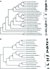
Figure 1
Figure 1. ACE and ACE2 inferred phylogeny of selected representative species
Protein sequences were aligned and analysed using Maximum Likelihood (ML) with 1000 replicates in MEGA X. Accession numbers were included with each taxon and branch supports are shown at each node indicating percentage agreement of node position amongst bootstrap replicates. Branch supports below 70% are not well supported. Animal icons were obtained through BioRender. Only metallopeptidase ACE homologues were included in the analysis and only incomplete sequences for Locusta migratoria, Chelonus inanitus and Myotis daubentonii were available at the time of sequence acquisition. (A) Invertebrate ACE and ACE-like peptides identified in venom are clustered together and included in the representative ACE phylogeny. Moreover, vertebrate mammalian ACE has greater evolutionary support of a close relationship than seen in the invertebrate species. (B) ACE2 phylogeny includes sequences of pangolin and bat species indicated as potential hosts for the SARS-CoV-2 virus. In some nodes, ACE2 may be too divergent due to missing data, gene flow or recombination for higher branch support values.
Figure 2
Figure 2. Schematic structure representations
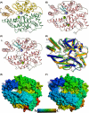
Crystal structures of (A) typical closed ACE N-domain (PDB code: 6F9V), (B) typical closed ACE C-domain (PDB code: 6H5W), (C) closed ACE2 (PDB code: 1R4L). (D) Overlay of ACE N- and C-domains with ACE2 ACE domain (blue, orange and green, respectively, with active site zinc ions shown as lighter coloured spheres). (E,F) Surface view of the ACE N-domain (PDB code: 6ZPQ) and ACE2 open structures (PDB code: 1R42), respectively. (A) shows subdomain 1 and 2 in orange (the lid-like region in dark orange) and green colour, respectively. The chloride and active site zinc ions are depicted as green and lilac spheres, respectively. For clearer comparison panels (B,C) have been coloured by secondary structure with α-helices and β-strands coloured rose and dark cyan, respectively.
Figure 3
Figure 3. Variation in subsites between ACE and ACE2
Panel (A) depicts the relevant residues in the S2′-S1 subsites with ACE2, N- and C-domains coloured green, blue and orange, respectively. The dotted arrow indicates that ACE2 S1′ Arg273 is equivalent to an S2′ residue in ACE. Binding cavity of (B) ACE2 in complex with MLN-4760 (PDB code: 1R4L) and (C) ACE C-domain in complex with omapatrilat (PDB code: 6H5W) showing variation in available space within the S2′-S1 subsites.
Figure 4
Figure 4. Mechanism of ACE domain opening
Overlay of the open and closed ACE2 structures highlighting the regions that move during the domain opening with the hinges about which the regions pivot numbered. Each moving region is also shown in isolation for clarity with the largest movement point marked. Subdomain 2 is shown in dark green/light green, with the moving regions of subdomain 1 coloured dark red/coral (residues 21–102), orange/yellow (residues 286–397), magenta/light pink (residues 397–431), dark blue/light blue (residues 521–561) and dark grey/light grey (residues 566–580). Zinc ion is shown as violet/lilac sphere. Darker colours are the closed structure.
Figure 5
Figure 5. ACE2 structures in complex with coronavirus RBDs
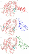
Schematic representations of ACE2 in complex with RBDs from (A) NL63-CoV (PDB code: 3KBH), (B) SARS-CoV (PDB code: 2AJF) and (C) SARS-CoV-2 (PDB code: 6M0J). ACE2 domains are coloured by secondary structure with α-helices and β-strands coloured rose and dark cyan, respectively. NL63-CoV, SARS-CoV and SARS-CoV-2 RBDs are coloured red, blue and green, respectively.

References

    1. Skeggs L.T. Jr, Kahn J.R. and Shumway N.P. (1956) The preparation and function of the hypertensin-converting enzyme. J. Exp. Med. 103, 295–299 10.1084/jem.103.3.295 - DOI - PMC - PubMed
    1. Kessler S.P., Rowe T.M., Gomos J.B., Kessler P.M. and Sen G.C. (2000) Physiological non- equivalence of the two isoforms of angiotensin-converting enzyme. J. Biol. Chem. 275, 26259–26264 10.1074/jbc.M004006200 - DOI - PubMed
    1. Hagaman J.R., Moyer J.S., Bachman E.S., Sibony M., Magyar P.L., Welch J.E. et al. . (1998) Angiotensin-converting enzyme and male fertility. Proc. Natl. Acad. Sci. U.S.A. 95, 2552–2557 10.1073/pnas.95.5.2552 - DOI - PMC - PubMed
    1. Erdos E.G. (1990) Angiotensin-I converting enzyme and the changes in our concepts through the years - Dahl, Lewis, K. Memorial Lecture. Hypertension. 16, 363–370 10.1161/01.HYP.16.4.363 - DOI - PubMed
    1. Ehlers M.R., Gordon K., Schwager S.L. and Sturrock E.D. (2012) Shedding the load of hypertension: the proteolytic processing of angiotensin-converting enzyme. S. Afr. Med. J. 102, 461–464 10.7196/SAMJ.5596 - DOI - PubMed

Publication types

MeSH terms