. 2021 Jan 6;179(1):135-146.
doi: 10.1093/toxsci/kfaa165.

Tungsten Increases Sex-Specific Osteoclast Differentiation in Murine Bone

Affiliations

Tungsten Increases Sex-Specific Osteoclast Differentiation in Murine Bone

Hsiang Chou et al. Toxicol Sci. .

Abstract

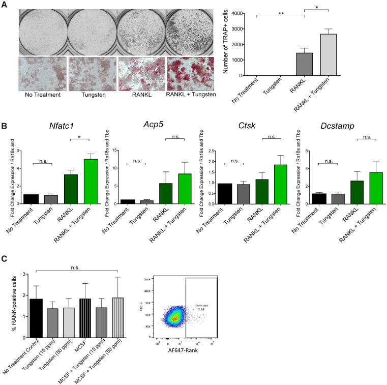
Tungsten is a naturally occurring metal that is increasingly used in industry and medical devices, and is labeled as an emerging environmental contaminant. Like many metals, tungsten accumulates in bone. Our previous data indicate that tungsten decreases differentiation of osteoblasts, bone-forming cells. Herein, we explored the impact of tungsten on osteoclast differentiation, which function in bone resorption. We observed significantly elevated osteoclast numbers in the trabecular bone of femurs following oral exposure to tungsten in male, but not female mice. In order to explore the mechanism(s) by which tungsten increases osteoclast number, we utilized in vitro murine primary and cell line pre-osteoclast models. Although tungsten did not alter the adhesion of osteoclasts to the extracellular matrix protein, vitronectin, we did observe that tungsten enhanced RANKL-induced differentiation into tartrate-resistant acid phosphatase (TRAP)-positive mononucleated osteoclasts. Importantly, tungsten alone had no effect on differentiation or on the number of multinucleated TRAP-positive osteoclasts. Enhanced RANKL-induced differentiation correlated with increased gene expression of differentiated osteoclast markers Nfatc1, Acp5, and Ctsk. Although tungsten did not alter the RANK surface receptor expression, it did modulate its downstream signaling. Co-exposure of tungsten and RANKL resulted in sustained positive p38 signaling. These findings demonstrate that tungsten enhances sex-specific osteoclast differentiation, and together with previous findings of decreased osteoblastogenesis, implicate tungsten as a modulator of bone homeostasis.

Keywords: RANKL; bone; differentiation; metals; mice; osteoclastogenesis; osteoclasts; signaling; trabecular bone; tungsten.


Figures

Figure 1.
Figure 1.
Tungsten increases the number of TRAP-positive osteoclasts in the trabecular region of femurs in male C57BL/6 mice. Femurs from C57BL/6 male and female mice exposed to 15 ppm tungsten or tap water for 4 weeks were sectioned and stained for osteoclastic marker TRAP. A, Representative images of TRAP-positive osteoclasts, stained red, and shown with black arrows, whereas the spongy bone is stained green. Images were taken at 10× and scale bar represents 100 μm. B, Total osteoclasts observed in the trabecular region of the femur sections. Graphs show the average number of osteoclasts and average osteoclast/area of bone marrow ± SEM, n = 4. Two-way ANOVA was carried out for statistics, *p < .05.
Figure 2.
Figure 2.
Tungsten does not affect adhesion of osteoclasts to vitronectin surfaces. A, Bone marrow-derived osteoclasts were differentiated on bacterial plates for 7 days and then re-seeded at 50 000 cells per well in a 24-well plate previously coated with vitronectin the night before. Cells were incubated for 45 min before washed off and TRAP stained. Four representative pictures were taken per well, summed, and average across 3 technical replicates per biological replicate. Graph shows the mean percent increase normalized to the RANKL treatment group ± SEM. n = 3. B, Expression of Itgav and Itgb3 integrin mRNA levels were assessed in M-CSF-induced osteoclasts exposed to 15 ppm tungsten, 15 ng/ml RANKL, or the combination. n = 3. *p < .05.
Figure 3.
Figure 3.
Tungsten enhances the number of RANKL-induced osteoclasts in vitro. Osteoclastogenesis was induced in primary bone marrow cells with 50 ng/ml MCSF and 15 or 50 ng/ml RANKL with or without 15 μg/ml tungsten. The total number of TRAP+ osteoclasts (A), as well as the total number and area of multinucleated osteoclasts (B) were enumerated. Representative images are shown, where scale bar represents 200 μm. Images (10×) were analyzed by taking the sum from 4 representative photos of each well and averaging across 4 technical replicates of each condition. The average area of multinucleated TRAP-positive osteoclasts was analyzed by measuring the size of each multinucleated osteoclast divided by the total number of TRAP-positive multinucleated osteoclasts. Graphs represent the mean ± SEM, n = 3. C, Bone marrow cells were exposed for 24 h to 15 or 50 μg/ml tungsten and the RANK expression on CD11b+ cells assessed by flow cytometry. n = 2. *p < .05; **p < .01; ***p < .001.
Figure 4.
Figure 4.
Tungsten enhances RANKL-induced osteoclastic gene markers in primary osteoclasts in vitro. Gene expression of osteoclast-specific genes (Nfatc1, Acp5, Ctsk, and Dcstamp) was quantified by qRT-PCR. Primary osteoclasts cultured in vitro in the presence of 50 ng/ml MCSF and 15 ng/ml RANKL with or without 15 ppm tungsten for 6 days, or in the presence of 50 ng/ml MCSF and 50 ng/ml RANKL with or without 15 ppm tungsten for 4 days. Graph shows the mean fold change in gene expression ± SEM. n = 3. *p < .05.
Figure 5.
Figure 5.
Tungsten increases the number of TRAP-positive osteoclasts in RAW 264.7 in vitro. A, RAW 264.7 were cultured with or without 15 μg/ml tungsten and with or without 15 ng/ml GST-sRANKL. After 5 days, positive TRAP staining of the total number of cells were counted using the GelCount plate scanner software. Graph shows the mean number for TRAP+ cells ± SEM, n = 4. Gray-scale pictures show representative well of each condition taken using the GelCount plate scanner and microscopic pictures are at the bottom showing the red TRAP stain. B, Gene expression of Nfatc1, Acp5, Ctsk, and Dcstamp were assessed after 24 h. Graph shows the mean ± SEM, n = 3, except for Nfatc1, n = 4. C, Cells were stained after 24 h and the RANK expression on CD11b+ cells assessed. n = 2. *p < .05; **p < .01.
Figure 6.
Figure 6.
Tungsten alters signaling downstream of RANK-RANKL. A, RAW 264.7 were cultured in 2.5% serum with or without 15 μg/ml tungsten 24 h after seeding, then induced with 15 ng/ml GST-sRANKL with or without 15 ppm tungsten and harvested over time. Whole cell extracts were used for immunoblots to detect levels of phospho- and total p38, phospho- and total ERK, phospho- and total JNK, phospho- and total p65, and phospho- and total mTOR. Representative blot for loading control of B-ACTIN is also shown. Numbers above phospho-blots indicate average relative densitometry values of phospho-protein/total protein/B-ACTIN across at least 3 different replicates. B, COS-1 cells were transiently transfected with an NF-κB-driven luciferase plasmid. Cells were treated with tungsten alone for 5 or 21 h or pretreated with tungsten and 20 ng/ml TNFα for the final 3 or 5 h. n = 3. *p < .05; **p < .01; ***p < .001; ****p < .0001.

References

    1. Agency for Toxic Substances and Disease Registry (ATSDR). (2005). Toxicological Profile for Tungsten ATSDR, Atlanta, GA. Available at: www.atsdr.cdc.gov/toxprofiles/tp186.pdf. Accessed November 4, 2020. - PubMed
    1. Beier E. E., Holz J. D., Sheu T. J., Puzas J. E. (2016). Elevated lifetime lead exposure impedes osteoclast activity and produces an increase in bone mass in adolescent mice. Toxicol. Sci. 149, 277–288. - PMC - PubMed
    1. Beier E. E., Maher J. R., Sheu T. J., Cory-Slechta D. A., Berger A. J., Zuscik M. J., Puzas J. E. (2013). Heavy metal lead exposure, osteoporotic-like phenotype in an animal model, and depression of wnt signaling. Environ. Health Perspect. 121, 97–104. - PMC - PubMed
    1. Bolt A. M., Grant M. P., Wu T. H., Flores Molina M., Plourde D., Kelly A. D. R., Negro Silva L. F., Lemaire M., Schlezinger J. J., Mwale F., et al. (2016). Tungsten promotes sex-specific adipogenesis in the bone by altering differentiation of bone marrow-resident mesenchymal stromal cells. Toxicol. Sci. 150, 333–346. - PMC - PubMed
    1. Bolt A. M., Mann K. K. (2016). Tungsten: An emerging toxicant, alone or in combination. Curr. Environ. Health Rep. 3, 405–415. - PubMed

Publication types

Substances

LinkOut - more resources