. 2020 Dec 24;63(24):15075-15093.
doi: 10.1021/acs.jmedchem.0c00812. Epub 2020 Nov 4.

Dienone Compounds: Targets and Pharmacological Responses

Affiliations

Dienone Compounds: Targets and Pharmacological Responses

Martina Bazzaro et al. J Med Chem. .

Abstract

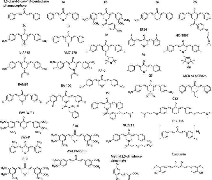
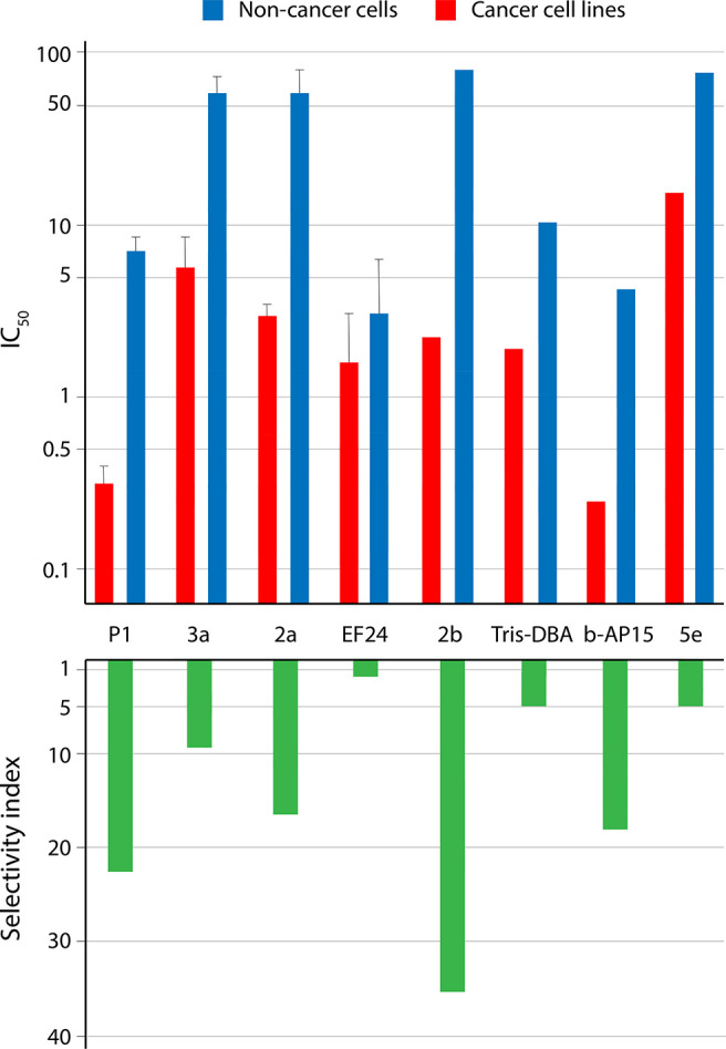
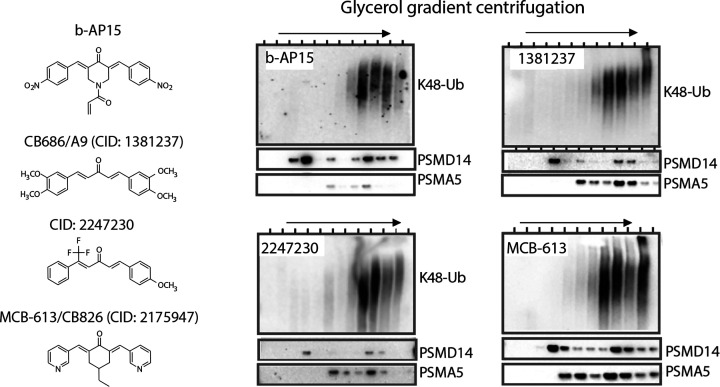
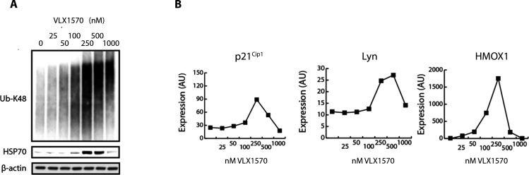
The biological responses to dienone compounds with a 1,5-diaryl-3-oxo-1,4-pentadienyl pharmacophore have been studied extensively. Despite their expected general thiol reactivity, these compounds display considerable degrees of tumor cell selectivity. Here we review in vitro and preclinical studies of dienone compounds including b-AP15, VLX1570, RA-9, RA-190, EF24, HO-3867, and MCB-613. A common property of these compounds is their targeting of the ubiquitin-proteasome system (UPS), known to be essential for the viability of tumor cells. Gene expression profiling experiments have shown induction of responses characteristic of UPS inhibition, and experiments using cellular reporter proteins have shown that proteasome inhibition is associated with cell death. Other mechanisms of action such as reactivation of mutant p53, stimulation of steroid receptor coactivators, and induction of protein cross-linking have also been described. Although unsuitable as biological probes due to widespread reactivity, dienone compounds are cytotoxic to apoptosis-resistant tumor cells and show activity in animal tumor models.


Conflict of interest statement

The authors declare no competing financial interest.

Figures

Figure 1
Figure 1
Chemical structures of some of the compounds discussed in the text.
Figure 2
Figure 2
Tumor cell selectivity of dienone compounds. The log10 IC50 values are shown on the y-axis in the upper graph and selectivity indices on the y-axis in the lower graph. Shown are the mean ± SD values where possible to calculate (i.e., where data from three or more cell lines were reported). The figure is based on the data in presented Supplementary Table 1.
Figure 3
Figure 3
b-AP15 and structurally related compound induce accumulation of polyubiquitinated proteins on proteasomes. Lysates from cells exposed to different compounds were fractionated by glycerol gradient centrifugation, and fractions were collected and processed for immunoblotting. Compounds CID1381237, CID2247230, and CID2175947 were identified in a cell-based screen for compounds stimulate accumulation of UbG76V-YFP in exposed cells. CID 2175947 (CB826 in ref (57)) is also known as MCB-613 and has been described to stimulate steroid receptor coactivator activity and to be preferentially cytotoxic to cancer cell lines. CID1381237 is also known as A9 (Figure 1) and C8. Data reproduced from ref (57), which licensed under CC BY 4.0.
Figure 4
Figure 4
Concentration-dependent responses to VLX1570. (A) Induction of polyubiquitinated proteins in RS4;11 ALL cells. Note strong increases of polyubiquitin and induction of Hsp70 at 250 nM VLX1570. (B) Increases in p21Cip1, LYN, and HMOX1. RS4;11 cells were exposed to VLX1570, and different markers were analyzed in the same extracts using a PCR-based multiplex method (OLINK, Uppsala, Sweden). Note the increases of HMOX1 at 50–100 nM VLX1570 and the decreases of all markers at high concentrations. Figure from ref (157), which licensed under CC BY 3.0.
Figure 5
Figure 5
Association between polyubiquitin association and cell death. (A) HCT116 colon cancer cells were exposed to 1 μM b-AP15 or 100 nM bortezomib and harvested at the indicated time points. Lysates were subjected to immunoblotting for K48-linked polyubiquitin, HSP-70B′, p21Cip1, PARP, or β-actin (loading control). (B) HCT116 cells or hTERT-RPE1 cells (immortalized epithelial cells) were exposed to different concentrations of b-AP15 for 1 h, followed by washing and incubation for 16 h in drug-free medium. Lysates were subjected to immunoblotting for K48-linked polyubiquitin, PARP and β-actin (loading control). Note the stronger accumulation of polyubiquitinated proteins and the induction of PARP cleavage in the colon cancer cells. (C) CpdA is an inhibitor of the Sec61 complex of anterograde protein export into the ER. Immortalized hTERT-RPE1 epithelial cells were exposed to increasing concentration of b-AP15 in the presence or absence of CpdA (“Sec61 inhib”). Note the increase in polyubiquitin accumulation, PARP cleavage, and cell death in the presence of CpdA (* p < 0.05). Parts A–C are reproduced from Sun et al., which is licensed under CC BY 4.0. (D) Graphic illustration of the results. Shown are proteasomes (red, 20S; green, 19S) and polyubiquitinated proteins.
Figure 6
Figure 6
Proteasome inhibition induced by b-AP15 is associated with cell death. (A) A reporter protein (UbG76V-YFP) is degraded by the proteasome (PS) in human melanoma cells. Proteasome inhibition (PI) leads to accumulation of the reporter protein in the cells. (B) Tracing individual cells expressing UbG76V-YFP following exposure to 250 nM b-AP15. Cells become UbG76V-YFP positive after 1–4 h, showing accumulation of the proteasome reporter. YFP-positive cells subsequently round up and the YFP leaks out from the cells after membrane disintegration. In contrast, cells that remain YFP-negative at 4 h of exposure survive and continue to divide. Results were obtained using an IncuCyte instrument (see refs (87) and (98)). (C) Time-lapse recordings of UbG76V-YFP-expressing human melanoma cells after addition of compounds. Upper panel: Human melanoma cells expressing UbG76V-YFP were exposed to 100 nM bortezomib and traced for 10 h. Notice the appearance of YFP fluorescence in cell nuclei at 2 h and later appearance in the cytoplasm (one cell highlighted at 2 h). Middle panel: Cells were exposed to 250 nM b-AP15. Note the similar pattern of initial nuclear and later cytoplasmic fluorescence (one cell highlighted at 1 h), similar to the pattern observed using bortezomib. Also note that cells either become positive and die or remain completely negative and survive. Lower panel, left: control. Vehicle-treated control cells remain YFP-negative and proliferate. Lower panel, right: >90% of the cells exposed to 500 nM b-AP15 become YFP-positive after 6 h, and most of these cells have rounded up at 18 h, some have lost membrane integrity and the YFP content. Original data were published in refs (87) and (98).
Figure 7
Figure 7
(A) Target affinity does not necessarily correlate to mechanism of action. A major fraction of cellular genes (∼90%) have been found to be nonessential for proliferation of tumor cells in vitro. Small molecules that bind to the protein products of these genes are therefore unlikely to affect tumor cell proliferation. In contrast, low affinity binding to essential proteins may result in strong inhibition of proliferation. An example is microtubule dynamics, known to be sensitive to low concentrations of small molecules and for which off-target effects may occur for various compounds when used in screens at 5–10 μM. (B) Illustration of responses to VLX1570 reported in different studies.,, HMOX1 is induced at lower concentration of b-AP15/VLX1570 than other markers (see Figure 4). Nrf-2/KEAP is targeted by electrophiles, and the Nrf-2 system is likely to be particularly sensitive to electrophiles. Increases of polyubiquitinated proteins, Hsp70 chaperones, and proteasome substrates such p21Cip1 are observed at somewhat higher concentrations of VLX1570; increased levels of Hsp70 are also observed at these concentrations (Figure 4). Inhibition of translation is observed at increasing concentrations of VLX1570,, leading to a blunted response of Hsp70, HMOX1, p21Cip1, and LYN expression (Figure 4). Depletion of anamorsin/CIAPIN1 was reported to occur at concentrations of 0.5–1 μM  and the formation of higher molecular weight protein complexes at 10 μM  (using HeLa cells).

Similar articles

Cited by

References

    1. Swinney D. C.; Anthony J. How were new medicines discovered?. Nat. Rev. Drug Discovery 2011, 10, 507–519. 10.1038/nrd3480. - DOI - PubMed
    1. Mayr L. M.; Bojanic D. Novel trends in high-throughput screening. Curr. Opin. Pharmacol. 2009, 9, 580–588. 10.1016/j.coph.2009.08.004. - DOI - PubMed
    1. Schenone M.; Dancik V.; Wagner B. K.; Clemons P. A. Target identification and mechanism of action in chemical biology and drug discovery. Nat. Chem. Biol. 2013, 9, 232–240. 10.1038/nchembio.1199. - DOI - PMC - PubMed
    1. Lamb J.; Crawford E. D.; Peck D.; Modell J. W.; Blat I. C.; Wrobel M. J.; Lerner J.; Brunet J. P.; Subramanian A.; Ross K. N.; Reich M.; Hieronymus H.; Wei G.; Armstrong S. A.; Haggarty S. J.; Clemons P. A.; Wei R.; Carr S. A.; Lander E. S.; Golub T. R. The connectivity map: using gene-expression signatures to connect small molecules, genes, and disease. Science 2006, 313, 1929–1935. 10.1126/science.1132939. - DOI - PubMed
    1. Jafari R.; Almqvist H.; Axelsson H.; Ignatushchenko M.; Lundback T.; Nordlund P.; Martinez Molina D. The cellular thermal shift assay for evaluating drug target interactions in cells. Nat. Protoc. 2014, 9, 2100–2122. 10.1038/nprot.2014.138. - DOI - PubMed

Publication types

MeSH terms