

Effect of PET-CT misalignment on the quantitative accuracy of cardiac 15O-water PET
- PMID: 33146863
- PMCID: PMC9163113
- DOI: 10.1007/s12350-020-02408-6
Effect of PET-CT misalignment on the quantitative accuracy of cardiac 15O-water PET
Abstract
Background: Quantification of myocardial blood flow (MBF) with PET requires accurate attenuation correction, which is performed using a separate CT. Misalignment between PET and CT scans has been reported to be a common problem. The purpose of the present study was to assess the effect of PET CT misalignment on the quantitative accuracy of cardiac 15O-water PET.
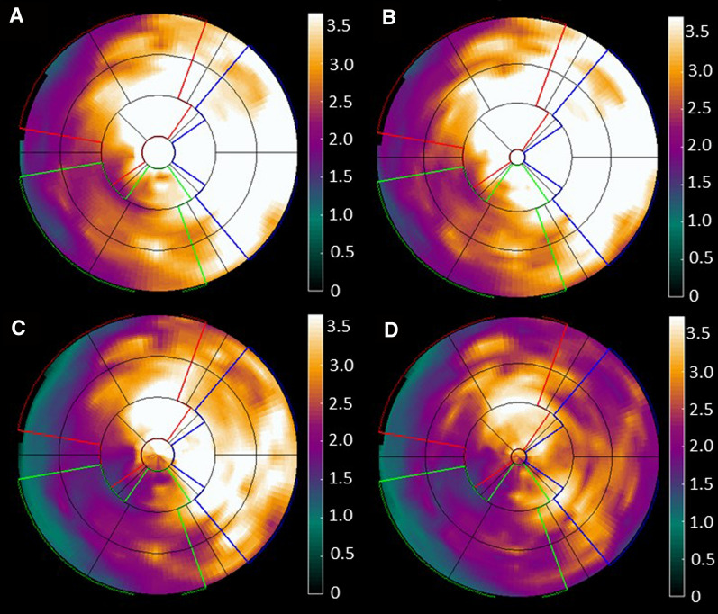
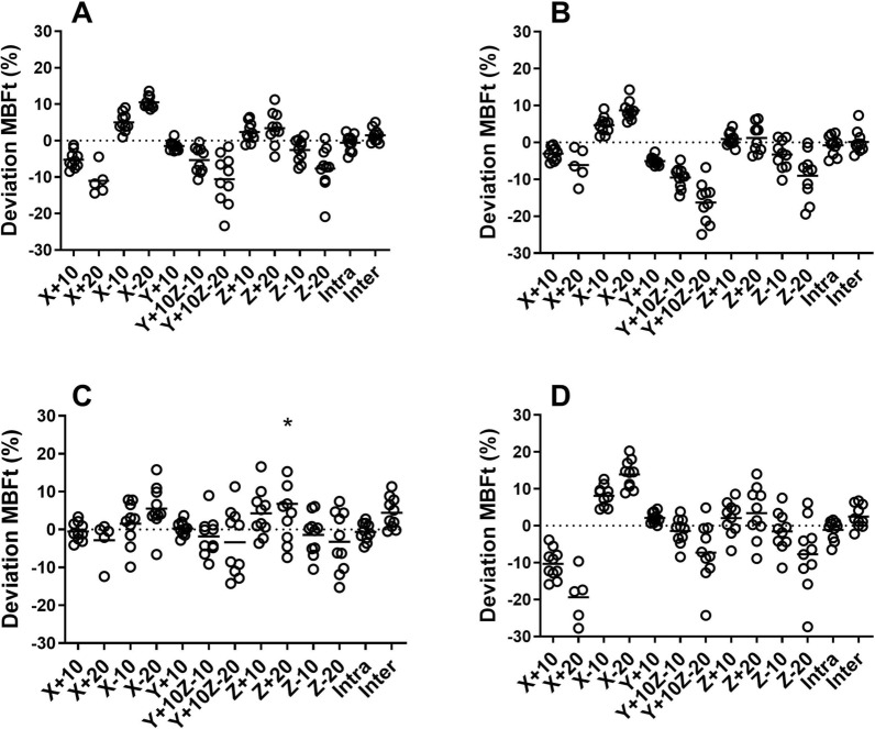
Methods: Ten clinical patients referred for evaluation of ischemia and assessment of MBF with 15O-water were included in the study. Eleven different misalignments between PET and CT were induced in 6 different directions with 10 and 20 mm amplitudes: caudal (+Z), cranial (- Z), lateral (±X), anterior (+Y), and anterior combined with cranial (+ Y and - Z). Blood flow was quantified from rates of washout (MBF) and uptake (transmural MBF, MBFt) for the whole left ventricle and the three coronary territories. The results from all misalignments were compared to the original scan without misalignment.
Results: MBF was only minorly affected by misalignments, but larger effects were seen in MBFt. On the global level, average absolute deviation across all misalignments for MBF was 1.7% ± 1.4% and for MBFt 5.4% ± 3.2 Largest deviation for MBF was - 4.8% ± 5.8% (LCX, X + 20) and for MBFt - 19.3% ± 9.6% (LCX, X + 20). In general, larger effects were seen in LAD and LCX compared to in RCA.
Conclusion: The quantitative accuracy of MBF from 15O-water PET, based on the washout of the tracer, is only to a minor extent affected by misalignment between PET and CT.
Keywords: PET; image analysis; myocardial blood flow; perfusion agents.
© 2020. The Author(s).
Figures


Comment in
-
Getting the right patient to angiography: Can we level the playing field?J Nucl Cardiol. 2022 Jun;29(3):1156-1158. doi: 10.1007/s12350-020-02475-9. Epub 2021 Jan 7. J Nucl Cardiol. 2022. PMID: 33415644 No abstract available.
-
Cardiac 15O-water PET: Does mismatched attenuation correction not matter?J Nucl Cardiol. 2022 Jun;29(3):1129-1131. doi: 10.1007/s12350-021-02573-2. Epub 2021 Mar 9. J Nucl Cardiol. 2022. PMID: 33751477 No abstract available.
References
-
- Danad I, Raijmakers PG, Driessen RS, Leipsic J, Raju R, Naoum C, Knuuti J, Maki M, Underwood RS, Min JK, Elmore K, Stuijfzand WJ, van Royen N, Tulevski II, Somsen AG, Huisman MC, van Lingen AA, Heymans MW, van de Ven PM, van Kuijk C, Lammertsma AA, van Rossum AC, Knaapen P. Comparison of coronary CT angiography, SPECT, PET, and hybrid imaging for diagnosis of ischemic heart disease determined by fractional flow reserve. JAMA Cardiol. 2017;2:1100–1107. doi: 10.1001/jamacardio.2017.2471. - DOI - PMC - PubMed
-
- Danad I, Uusitalo V, Kero T, Saraste A, Raijmakers PG, Lammertsma AA, Heymans MW, Kajander SA, Pietila M, James S, Sorensen J, Knaapen P, Knuuti J. Quantitative assessment of myocardial perfusion in the detection of significant coronary artery disease: Cutoff values and diagnostic accuracy of quantitative [(15)O]H2O PET imaging. J Am Coll Cardiol. 2014;64:1464–1475. doi: 10.1016/j.jacc.2014.05.069. - DOI - PubMed
-
- Kajander S, Joutsiniemi E, Saraste M, Pietila M, Ukkonen H, Saraste A, Sipila HT, Teras M, Maki M, Airaksinen J, Hartiala J, Knuuti J. Cardiac positron emission tomography/computed tomography imaging accurately detects anatomically and functionally significant coronary artery disease. Circulation. 2010;122:603–613. doi: 10.1161/CIRCULATIONAHA.109.915009. - DOI - PubMed
MeSH terms
Substances
LinkOut - more resources
Full Text Sources
