. 2020 Dec 15;31(26):2932-2947.
doi: 10.1091/mbc.E19-10-0591. Epub 2020 Nov 4.

PTP-3 phosphatase promotes intramolecular folding of SYD-2 to inactivate kinesin-3 UNC-104 in neurons

Affiliations

PTP-3 phosphatase promotes intramolecular folding of SYD-2 to inactivate kinesin-3 UNC-104 in neurons

Muthaiyan Shanmugam Muniesh et al. Mol Biol Cell. .

Abstract

UNC-104 is the Caenorhabditis elegans homolog of kinesin-3 KIF1A known for its fast shuffling of synaptic vesicle protein transport vesicles in axons. SYD-2 is the homolog of liprin-α in C. elegans known to activate UNC-104; however, signals that trigger SYD-2 binding to the motor remain unknown. Because SYD-2 is a substrate of PTP-3/LAR PTPR, we speculate a role of this phosphatase in SYD-2-mediated motor activation. Indeed, coimmunoprecipitation assays revealed increased interaction between UNC-104 and SYD-2 in ptp-3 knockout worms. Intramolecular FRET analysis in living nematodes demonstrates that SYD-2 largely exists in an open conformation state in ptp-3 mutants. These assays also revealed that nonphosphorylatable SYD-2 (Y741F) exists predominately in folded conformations, while phosphomimicking SYD-2 (Y741E) primarily exists in open conformations. Increased UNC-104 motor clustering was observed along axons likely as a result of elevated SYD-2 scaffolding function in ptp-3 mutants. Also, both motor velocities as well as cargo transport speeds were visibly increased in neurons of ptp-3 mutants. Lastly, epistatic analysis revealed that PTP-3 is upstream of SYD-2 to regulate its intramolecular folding.


Figures
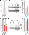
FIGURE 1:
FIGURE 1:
Changes in syd-2/unc-104 gene expression levels and SYD-2/UNC-104 protein interactions in ptp-3 mutants. (A, D) Relative quantification of mRNA expression levels of syd-2 and unc-104 from lysates of young adult worms (N2 control, ptp-3(ok244) and ptp-3(mu256) allelic mutants). Quantitative PCR was performed thrice and each trial was performed in triplicates. (B, E) Upper lane: Western blot of pulled-down UNC-104::GFP using anti-GFP antibody. Bottom lane: Blot of co-IP SYD-2 detected using anti-SYD-2 polyclonal antibody from lysates of ok244 and mu256 mutants. (C, F) Quantification of coprecipitated SYD-2 (normalized with UNC-104::GFP) from different genetic backgrounds. Error bars: ± max. and min. range. Unpaired Student’s t test. *p < 0.05, **p < 0.01, ***p < 0.001; t, df = 1.126, 4 (A, syd-2); 0.7039, 4 (A, unc-104); 4.389, 4 (D, syd-2); 11, 4 (D, unc-104); 0.6547, 4 (C) and 7.182, 4 (F).
FIGURE 2:
FIGURE 2:
SYD-2/PTP-3B colocalization and BiFC analysis. (A–E) PTP-3B::CFP (pseudo-colored in magenta) and GFP::SYD-2 colocalization in nerve rings (A–D) and VNCs (E). (D) Pseudo-color PDM image (see Materials and Methods) indicating quantifiable colocalization between SYD-2 and PTP-3B in the nerve ring. (F) Pearson’s correlation coefficient quantification of data shown in C. (G) Positive PTP-3B/SYD-2 BiFC signal in the nerve ring of living worms indicating that SYD-2 and PTP-3B are at least 7–10 nm close to each other (see Materials and Methods). (H) Worm expressing SNB-1::mRFP under a pan-neuronal promoter. (I) Merged image from G and H. (J) Stacks of straightened VNCs and ALM neurons with positive PTP-3B/SYD-2 BiFC signals. *Location of somata in ALM neurons. Dotted, white line marks the contour of the worm head. Scale bars: 10 µm. N = 10 worms in F. Error bar: ± max. and min. range. A indicates the anterior direction and P the posterior direction. For BiFC-positive and -negative controls, refer to Supplemental Figure S3, C and D.
FIGURE 3:
FIGURE 3:
SYD-2 exists predominantly in an open conformation in ptp-3(mu256) mutants. (A) Pictorial representation of the designed intramolecular FRET assay for this study. (B) Images of C. elegans nerve rings from the respective fluorescence channel. (C) Quantifi­cation of FRET efficiencies of Cypet::SYD-2::Ypet in different genetic backgrounds, where ptp-3 RNAi, ptp-3(mu256) and phosphomi­micking SYD-2 Y741E all lead to a significant decrease in FRET ratios while the nonphosphorylatable SYD-2 Y741F reveals wild type signals. Scale bar: 10 µm. N = 26 images (C). Error bars: ± max. and min. range. One-way ANOVA with Fisher’s LSD test. *p < 0.05, **p < 0.01, ***p < 0.001, and ****p < 0.0001. F (DFn, DFd) = 15.83 (7, 200). A indicates anterior direction and P the posterior direction. Dotted, white line marks the contour of the worm head.
FIGURE 4:
FIGURE 4:
STVs accumulate in somata of ptp-3 mutants. (A) Straightened ALM neurons taken from different worms within a population and stacked on each other. (B–D) Quantification of size (B), densities (C), and travel distances (D) of SNB-1 containing synaptic vesicles from images displayed in A. (E) Cartoon of HSN neuron with its presynaptic area and representative images of SNB-1 cargo distribution in wild type and mutants. *Soma of neuron. White arrow indicates the location of the vulva. Green boxes indicate the region used for quantification of fluorescence intensity of axons (blue dashed line) and synapses (red circles). P indicates the posterior direction and A the anterior direction. (F) Representative images of VNC and DNC from different study groups, straightened and stacked. (G) Quantification (fluorescence intensity) of the amount of cargo in the cell body and the IAR of ALM, nerve cord (VNC somata and DNC synapses), and HSN neurons (soma, axon, and synapse). Scale bars: 10 µm. Box plot with median and error bars: ± max. and min. range. One-way ANOVA with Fisher’s LSD multiple comparison test. *p < 0.05 and **p < 0.01. F (DFn, DFd) = 3.426 (2, 320) (B); 3.862 (2, 27) (G, ALM); 4.327 (2, 43) (G, HSN soma); 2.455 (2, 43) (G, HSN axon) and 3.909 (2, 43) (G, HSN synapse).
FIGURE 5:
FIGURE 5:
UNC-104 clustering is increased in ptp-3 mutants and decreased in syd-2 mutants. (A) UNC-104::mRFP accumulations and distribution patterns in straightened segments of sublateral neurons taken from different genetic backgrounds stacked on each other with: control = unc-104(e1265) allelic mutants rescued with UNC-104::mRFP; ptp-3(ok244) = ok244 allelic mutants expressing UNC-104::mRFP; ptp-3(mu256) = mu256 allelic mutants expressing UNC-104::mRFP; ptp-3(mu256) rescue = mu256 allelic mutants expressing both PTP-3B::CFP and UNC-104::mRFP; syd-2(ok217) = ok217 allelic mutants expressing UNC-104::mRFP; syd-2(ok217);ptp-3(mu256) = allelic double mutants expressing UNC-104::mRFP; syd-2(ok217) rescue = ok217 allelic mutants expressing both GFP::SYD-2 and UNC-104::mRFP; syd-2(ok217)[SYD-2 Y741F] = ok217 allelic mutant expressing both nonphosphorylatable GFP::SYD-2 Y741F and UNC-104::mRFP; syd-2(ok217)[SYD-2 Y741E] = ok217 allelic mutant expressing both phosphomimicking GFP::SYD-2 Y741E and UNC-104::mRFP. (B) UNC-104 cluster sizes in sublateral neurons. (C) UNC-104 densities along sublateral neurons. (D) Representative immunoblots from co-IP of UNC-104 from phosphodeficient (Y741F) and phosphomimicking (Y741E) SYD-2. (E) Quantification of coprecipitated UNC-104 either with phosphodeficient or phosphomimicking SYD-2. Scale bar: 10 µm. Box plot with median and error bars: ± max. and min. range. One-way ANOVA with Fisher’s LSD multiple comparison test. *p < 0.05, **p < 0.005, ***p < 0.001, and ****p < 0.0001. F (DFn, DFd) = 7.073 (8, 238) (C); 6.543 (2,6) (E).
FIGURE 6:
FIGURE 6:
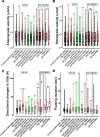
Both motor and cargo speeds are increased in ptp-3(mu256) mutants. (A) UNC-104 and SNB-1 anterograde velocities in various genetic backgrounds as well as after ptp-3 RNAi. (B) UNC-104 and SNB-1 retrograde velocities in various genetic backgrounds as well as after ptp-3 RNAi. (C) Quantification of motor and cargo reversals (number of directional changes within 100s). (D) Quantification of motor and cargo pausing duration. (E, F) Quantification of total anterograde (E) and (F) retrograde run length. (G) Quantification of net run length. Analyzed UNC-104 particles: > 1500 events, SNB-1 particles: > 750 events. See Supplemental Figure S5 for definition of motility parameters. Box plots with median and error bars: ± max. and min. range. One-way ANOVA with Fisher’s LSD multiple comparison test for UNC-104 in mutant strains and t test with Welch’s correction for RNAi and SNB-1. *p < 0.05, **p < 0.005, ***p < 0.001 and ****p < 0.0001. F (DFn, DFd) = 12.60 (7, 4346) (A); 3.175 (7, 4544) (B); 7.156 (7, 416) (C); 3.496 (7, 419) (D); 3.119 (7, 85) (E); 3.923 (7, 85) (F) and 3.035 (7, 97) (G).
FIGURE 7:
FIGURE 7:
PTP-3 regulates binding of SYD-2 to kinesin-3 UNC-104 to modulate fast axonal transport. From this study, it is evident that PTP-3 is upstream of SYD-2 to regulate UNC-104 activity. PTP-3 dephosphorylates SYD-2 at position Y741 resulting in intramolecular folding of SYD-2 concomitantly inactivating UNC-104. Lack of PTP-3 results in more open configurations of SYD-2 resulting in increased interactions with UNC-104 promoting increased UNC-104 scaffolding along axons and boosting speeds of present UNC-104 motors as well as STVs. Mild accumulation of STVs in the soma is likely the result of reduced gene expression of syd-2 and unc-104 in ptp-3 mutants.

Similar articles

Cited by

References

    1. Ackley BD, Harrington RJ, Hudson ML, Williams L, Kenyon CJ, Chisholm AD, Jin Y (2005). The two isoforms of the Caenorhabditis elegans leukocyte-common antigen related receptor tyrosine phosphatase PTP-3 function independently in axon guidance and synapse formation. J Neurosci 25, 7517–7528. - PMC - PubMed
    1. Anthis NJ, Haling JR, Oxley CL, Memo M, Wegener KL, Lim CJ, Ginsberg MH, Campbell ID (2009). Beta integrin tyrosine phosphorylation is a conserved mechanism for regulating talin-induced integrin activation. J Biol Chem 284, 36700–36710. - PMC - PubMed
    1. Blangy A, Lane HA, d’Herin P, Harper M, Kress M, Nigg EA (1995). Phosphorylation by p34cdc2 regulates spindle association of human Eg5, a kinesin-related motor essential for bipolar spindle formation in vivo. Cell 83, 1159–1169. - PubMed
    1. Brady ST, Morfini GA (2017). Regulation of motor proteins, axonal transport deficits and adult-onset neurodegenerative diseases. Neurobiol Dis 105, 273–282. - PMC - PubMed
    1. Brenner S (1974). The genetics of Caenorhabditis elegans. Genetics 77, 71–94. - PMC - PubMed

Publication types

MeSH terms

Substances

LinkOut - more resources