. 2020 Nov 4;15(11):e0240900.
doi: 10.1371/journal.pone.0240900. eCollection 2020.

Decorated bodies for eternal life: A multidisciplinary study of late Roman Period stucco-shrouded portrait mummies from Saqqara (Egypt)

Affiliations

Decorated bodies for eternal life: A multidisciplinary study of late Roman Period stucco-shrouded portrait mummies from Saqqara (Egypt)

Stephanie Zesch et al. PLoS One. .

Abstract

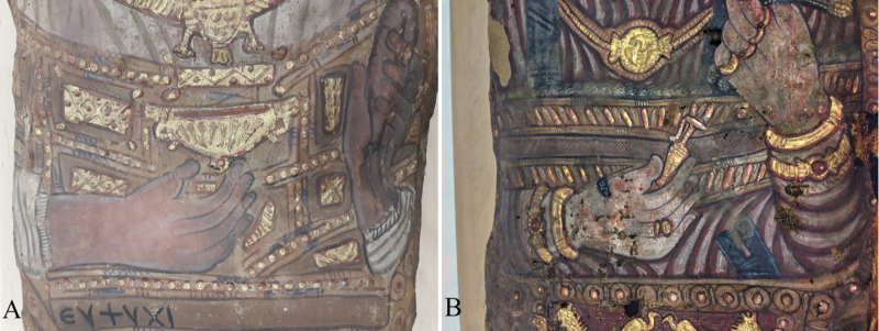
This study focuses on the multidisciplinary investigation of three stucco-shrouded mummies with mummy portrait from Egypt dating from the late 3rd to the middle of the 4th century AD, corresponding to the late Roman Period. These three mummies were excavated in the early 17th and late 19th centuries in the Saqqara necropolis near the ancient Egyptian capital of Memphis. Two of them experienced an interesting collection history, when they became part of the collection of the Elector of Saxony and King of Poland August II in Dresden, Germany, in 1728. The investigation includes information about the mummies' discovery, collection history and shroud decoration obtained through Egyptological expertise. In addition, information on the state of preservation, technique of artificial mummification, age at death, sex, body height and health of the deceased was achieved through computed tomography (CT) analysis. Research yielded an adult male, a middle-aged female and a young female. Due to the rather poorly preserved bodies of the male and middle-aged female, a specific technique of artificial mummification could not be ascertained. Brain and several internal organs of the well-preserved young female were identified. Wooden boards, beads of necklaces, a hairpin, and metal dense items, such as lead seals, nails and two coins or medallions were discovered. Paleopathological findings included carious lesions, Schmorl's nodes, evidence of arthritis and a vertebral hemangioma. The study revealed insights on the decoration and burial preparation of individuals of upper socioeconomic status living in the late Roman Period, as well as comprehensive bioanthropological information of the deceased.


Conflict of interest statement

One of the authors (WR) is affiliated to the Curt-Engelhorn-Center Archaeometry gGmbH. gGmbH is a non-profit company. There are no other relevant declarations relating to employment, consultancy, patents, products in development, or marketed products, etc. This does not alter our adherence to PLOS ONE policies on sharing data and materials.

Figures

Fig 1
Fig 1. Stucco-shrouded portrait mummies housed at the Dresden State Art Collections.
(A) male mummy (Aeg 777); (B) female mummy (Aeg 778) (© Sculpture Collection, Dresden State Art Collections, photos: H.-P. Klut/E. Estel).
Fig 2
Fig 2. Reproductions of copper engravings.
(A) a portrait of Pietro Della Valle; (B) the supposed moment of discovery of the mummies in Saqqara (adapted and reprinted under a CC BY license) [40].
Fig 3
Fig 3. Reproductions of copper engravings.
(A) a portrait of Athanasius Kircher made by Cornelis Bloemaert (original (inv. MP 12693) at Germanisches Nationalmuseum, Nuremberg, adapted and reprinted under a CC BY license); (B) the portrait mummies depicted by Athanasius Kircher (adapted and reprinted from [41] under a CC BY license).
Fig 4
Fig 4. The mummies’ acquisition for the art collection of the Elector of Saxony and King of Poland August II.
(A) a portrait of the sovereign made by Louis de Silvestre (original (inv. Gal.-Nr. 3943) at Old Masters Picture Gallery) (© Old Masters Picture Gallery, Dresden State Art Collections, photo: H.-P. Klut/E. Estel); (B) a copper engraving made by Anna Maria Werner and Christoph Raimund Thomann illustrating four mummies of the collection, including these two portrait mummies in the center (adapted and reprinted from [42] under a CC BY license).
Fig 5
Fig 5. Female stucco-shrouded portrait mummy (CG 33281) housed at the Museum of Egyptian Antiquities, Cairo.
© I. Badr.
Fig 6
Fig 6. Detail views of the mummy portraits and jewelry depicted on the shrouds.
(A) male mummy (Aeg 777); (B) female mummy (Aeg 778) (© Sculpture Collection, Dresden State Art Collections, photos: H.-P. Klut/E. Estel).
Fig 7
Fig 7. Detail views of the objects visible in the hands.
(A) male (Aeg 777) with a so-called kantharos in his right hand and a garland or a folded wreath of blossoms in his left hand. Note also the Greek inscription below his right forearm; (B) female (Aeg 778) with a so-called lekythos in her right hand. (© Sculpture Collection, Dresden State Art Collections, photos: M. Gander/M. Loth).
Fig 8
Fig 8. Young female mummy (CG 33281).
The detail photo focuses on the red-brown pointed shoes, surrounded by white seams on the front and sides, decorated with a circular object on each tip and two white stars in the center of each shoe (© I. Badr).
Fig 9
Fig 9. Young female mummy (CG 33281).
The photo shows the decoration of the mummy shroud, focusing on the portrait and torso (© I. Badr).
Fig 10
Fig 10. Male mummy (Aeg 777).
(A) thick-slab mean intensity coronal multi-planar reconstruction illustrates one metal object close to the right femur, suggested to be a translocated metal seal, and another metal seal on the outer textile layer (arrows); (B) sagittal multi-planar reconstruction visualizes parts of a broken wooden board placed beneath the body (arrows).
Fig 11
Fig 11. Male mummy (Aeg 777).
Thick-slab maximum intensity coronal multi-planar reconstruction illustrates disarticulated and partially broken bones of the trunk and the upper extremities, as well as a foreign object in the upper left thoracic region suggested to be a translocated metal seal. Scattered radiopaque material (possibly sediment and plant materials) are visible.
Fig 12
Fig 12. Female mummy (Aeg 778).
(A) thick-slab mean intensity coronal multi-planar reconstruction visualizes two circular metal dense foreign objects close to the right femur, probably representing coins or medallions. Two nails were identified in the abdominal region (arrow); (B) sagittal multi-planar reconstruction shows an intact wooden board identified beneath the body (arrow heads).
Fig 13
Fig 13. Young female mummy (CG 33281).
(A) thick-slab mean intensity coronal multi-planar reconstruction illustrates one of seven metal seals fixed on the outermost textile layer (arrow); (B) sagittal multi-planar reconstruction shows an intact wooden board placed beneath the body inside the wrappings (arrow heads).
Fig 14
Fig 14. Young female mummy (CG 33281).
Coronal multi-planar reconstruction depicts several segments of the innermost textile layer with a geometric pattern (asterisks) and longitudinal thin-walled low density beads in the thoracic region (arrows).
Fig 15
Fig 15. Young female mummy (CG 33281) with shrunken brain and brainstem preserved.
(A) sagittal multi-planar reconstruction; (B) coronal multi-planar reconstruction.
Fig 16
Fig 16. Male mummy (Aeg 777).
The detail photo shows a lead seal at the outermost textile layer on the left side of the mummy. The seal is fixed at the level of the mummy’s thigh with a cord about 15 cm in length. An almond-shaped depression in the center of the seal is most probably the insignia of the mummification workshop (© Sculpture Collection, Dresden State Art Collections, photos: M. Gander/M. Loth).
Fig 17
Fig 17. Female mummy (Aeg 778).
(A) volume rendered reconstruction illustrates numerous scattered beads in the thoracic region; (B) sagittal maximum intensity reconstruction shows a few of the perforated beads in detail (arrows).
Fig 18
Fig 18. Volume rendered reconstructions representing details of the young female mummy (CG 33281).
(A) numerous beads around the neck and on the thoracic region, suggesting an intact necklace/or several necklaces; (B) a hairpin on top of the head indicating an upswept hairstyle.
Fig 19
Fig 19. Skull of the male mummy (Aeg 777).
3D maximum intensity projection visualizes the unerupted permanent maxillary and mandibular left canines as a congenital dental anomaly.
Fig 20
Fig 20. Multi-planar reconstructions visualizing the knees of the female mummy (Aeg 778).
(A) both knees in axial view with evidence of juxtaarticular osteoporosis in the left knee; (B) the right knee joint in sagittal view without any pathological changes; (C) the left knee joint in sagittal view with evidence of arthritis, such as lucencies of the subchondral bone in the femorotibial and femoropatellar joints, and the patella subluxed inferiorly, contacting the proximal tibia.
Fig 21
Fig 21. Segment of the thoracic spine of the young female mummy (CG 33281).
Multi-planar reconstructions show a focal area of coarsening of the vertical trabeculae without expansion of the cortex, indicating a vertebral hemangioma of T8 thoracic vertebra in (A) sagittal, (B) axial, and (C) coronal views (arrow heads). Note a small-size osteoma (arrow) dorsally within the trabecular bone of T9 thoracic vertebra in (A).

Similar articles

Cited by

References

    1. Dannenfeldt KH. Egyptian Mumia: The Sixteenth Century Experience and Debate. Sixt Century J, 1985; 16(2): 163–180. https://www.jstor.org/stable/2540910 - PubMed
    1. Ikram S, Dodson A. The Mummy in Ancient Egypt: Equipping the Dead for Eternity. London: Thames and Hudson; 1998.
    1. Aufderheide AC. The Scientific Study of Mummies. Cambridge: Cambridge University Press; 2003.
    1. Rühli FJ, Chhem RK, Böni T. Diagnostic paleoradiology of mummified tissue: interpretation and pitfalls. Can Assoc Radiol J. 2004; 55(4): 218–227. - PubMed
    1. Hawass Z, Gad YZ, Ismail S, Khairat R, Fathalla D, Hasan N, et al. Ancestry and Pathology in King Tutankhamun’s Family. JAMA. 2010; 303(7): 638–647. http://jama.ama-assn.org/cgi/content/full/303/7/638 10.1001/jama.2010.121 - DOI - PubMed

Publication types