. 2021 Jan 6;109(1):59-72.e5.
doi: 10.1016/j.neuron.2020.10.012. Epub 2020 Nov 3.

Role of Aberrant Spontaneous Neurotransmission in SNAP25-Associated Encephalopathies

Affiliations

Role of Aberrant Spontaneous Neurotransmission in SNAP25-Associated Encephalopathies

Baris Alten et al. Neuron. .

Abstract

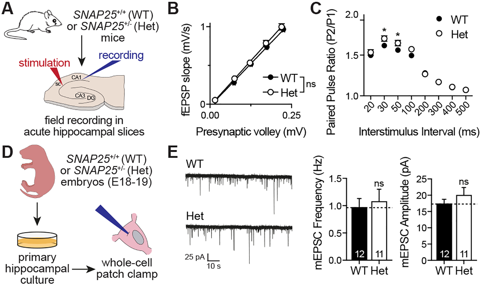
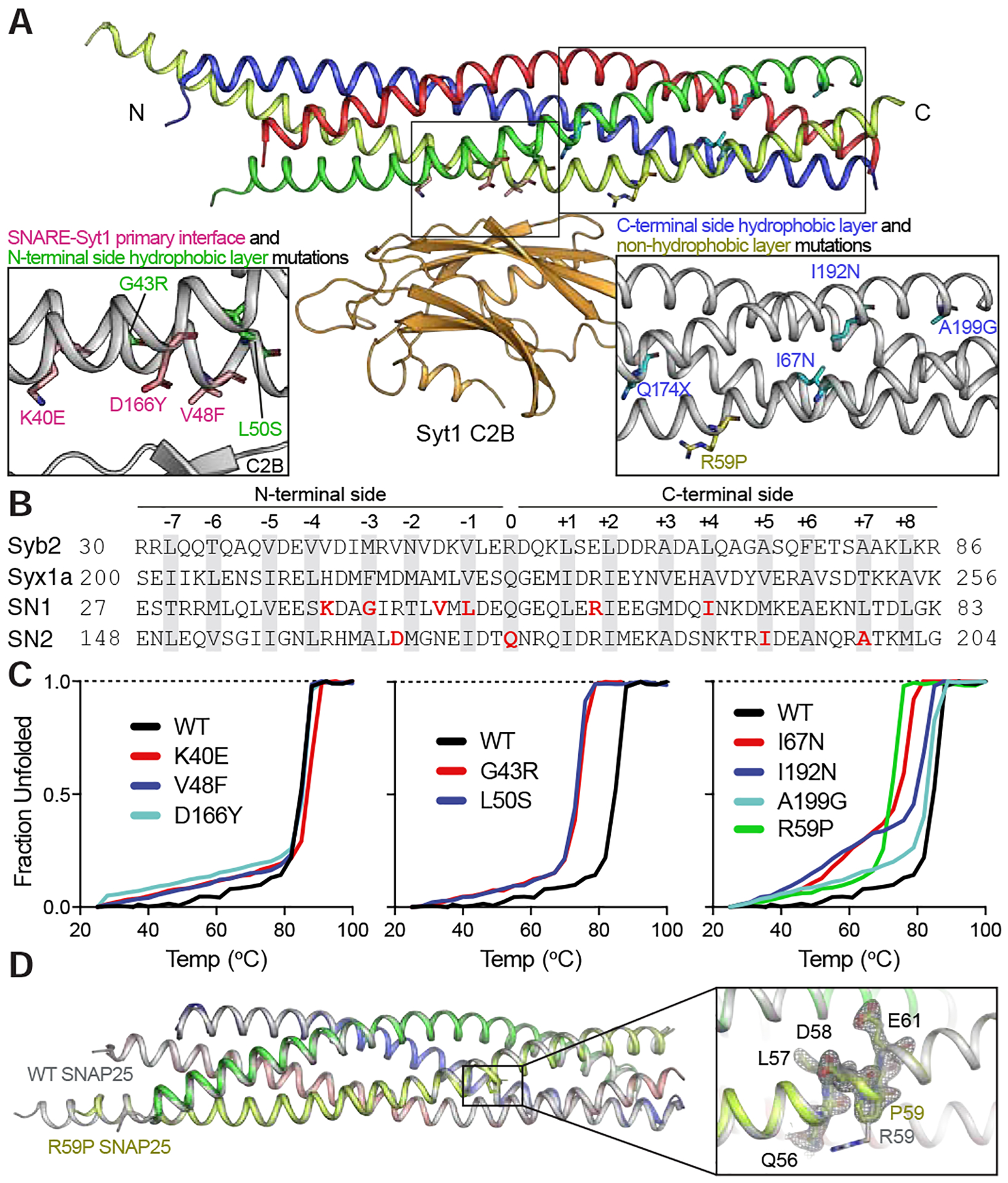
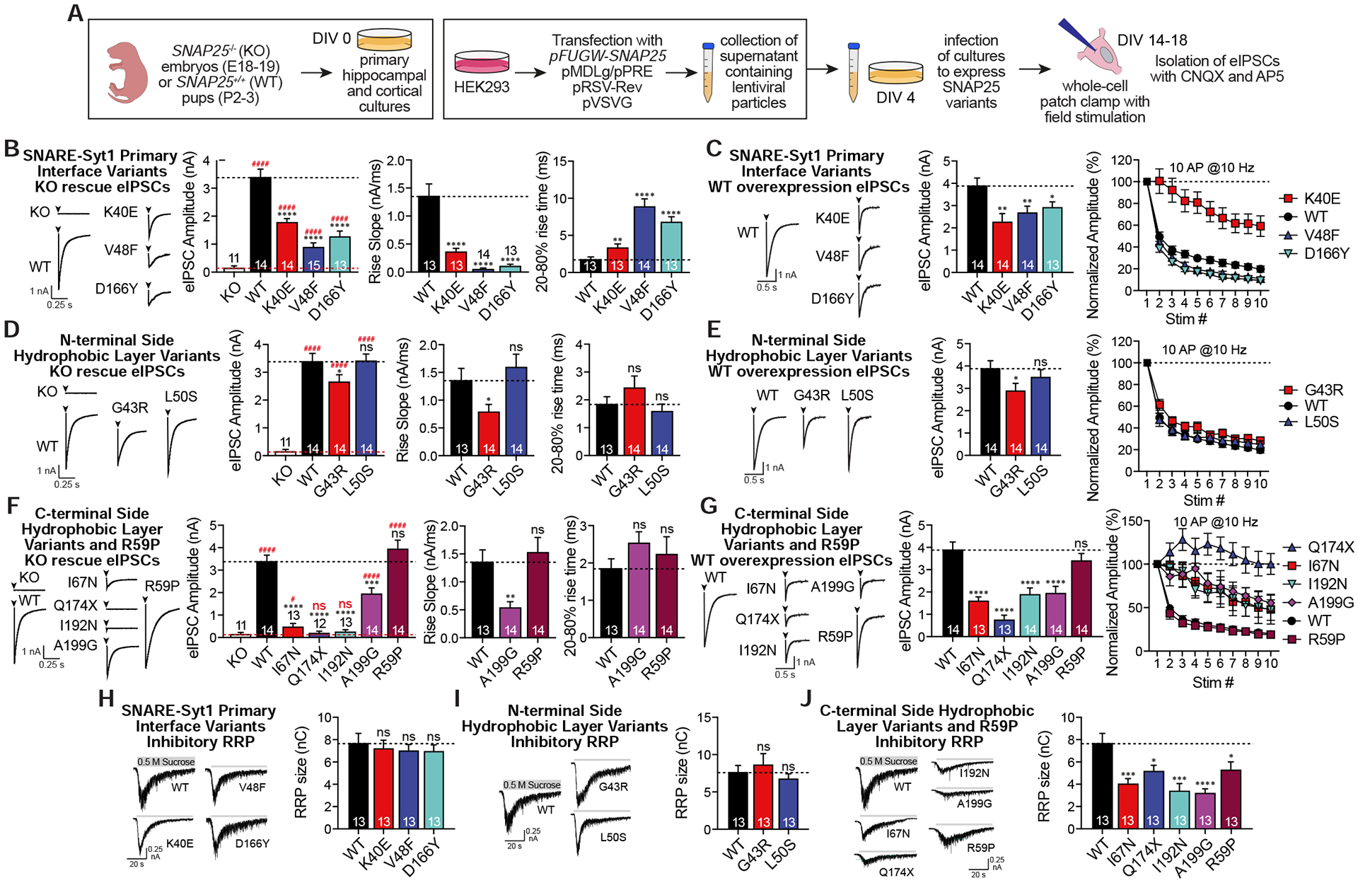
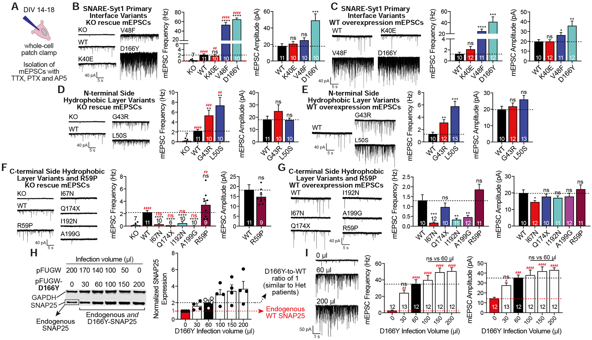
SNARE (soluble N-ethylmaleimide sensitive factor attachment protein receptor) complex, composed of synaptobrevin, syntaxin, and SNAP25, forms the essential fusion machinery for neurotransmitter release. Recent studies have reported several mutations in the gene encoding SNAP25 as a causative factor for developmental and epileptic encephalopathies of infancy and childhood with diverse clinical manifestations. However, it remains unclear how SNAP25 mutations give rise to these disorders. Here, we show that although structurally clustered mutations in SNAP25 give rise to related synaptic transmission phenotypes, specific alterations in spontaneous neurotransmitter release are a key factor to account for disease heterogeneity. Importantly, we identified a single mutation that augments spontaneous release without altering evoked release, suggesting that aberrant spontaneous release is sufficient to cause disease in humans.

Keywords: SNAP25; child neurology; development; developmental delay; epilepsy; exocytosis; neurotransmitter; spontaneous release; synaptic transmission; synaptotagmin.


Conflict of interest statement

Declaration of Interests The authors declare no competing interests.

Figures

Figure 1.
Figure 1.. Effects of SNAP25 haploinsufficiency on synaptic transmission.
A. Experimental design for acute hippocampal slice field electrophysiology. B. Input-output curves are not statistically different between SNAP25 Het mice and littermate WTs (n= 13 slices from 3 mice/group, slopes of the curves were compared using simple linear regression module of Prism 8, F1,253=2.819, p>0.05). C. Paired pulse ratio was increased in hippocampal slices of SNAP25 Het mice compared to littermate WTs (n= 13 slices from 3 mice/group, compared by multiple t-tests corrected for multiple comparisons using the Holm-Sidak method, p<0.05 for interstimulus intervals of 30 and 50 ms). D. Experimental design for whole cell patch clamp electrophysiology. E. Representative mEPSC traces with quantitative analysis of mEPSC frequencies and amplitudes.
Figure 2.
Figure 2.. Structural characterization of SNAP25 variants.
A. Structure of the SNARE complex and its primary interface with C2B domain of Syt1 (synaptobrevin-2 (syb2) blue, syntaxin-1a (syx1a) red, SNAP25 SN1 yellow, SNAP25 SN2 green, Syt1 C2B orange). Mutated residues are marked in insets for better visualization. B. Amino acid sequence map of individual SNARE motifs forming the ternary SNARE complex. 15 hydrophobic layers and the central ionic layer are highlighted in grey, whereas mutated SNAP25 residues are shown in red. C. Thermal melting curves using CD spectroscopy observed at 220 nm. D. Crystal structure of the ternary SNARE complex formed with the R59P SNAP25 variant (in color) superimposed on the complex formed with WT SNAP25 (in grey, PDB ID 1N7S). The omit map (mFo-DFc electron density map) of the residue region 56–61 from SNAP25 countered at 2.5 σ (grey mesh). See also supplementary table 1 and supplementary movies 1 and 2.
Figure 3.
Figure 3.. Effects of SNAP25 variants on evoked neurotransmission.
A. Experimental design. B-G. Representative eIPSC traces with the quantitative analysis of the amplitudes, rise slopes and 20–80% rise times of eIPSCs in response to single stimulation and eIPSC responses to repetitive 10 Hz 10 stimulations were presented for each group of SNAP25 variants expressed either on KO background or on WT background. SNAP25 variants were individually compared against either KO (red dashed line, significance denoted with #) or WT (black dashed line, significance denoted with *). Rise slopes and rise times of eIPSCs of the I67N, Q174X and I192N were not analysed since they are either not functional or nearly non-functional. H-J. Effects of SNAP25 variants overexpressed on WT cultures on the size of inhibitory readily releasable pool measured as a response to hypertonic sucrose perfusion.
Figure 4.
Figure 4.. Effects of SNAP25 variants on spontaneous neurotransmission.
A. Experimental design was similar to that of evoked neurotransmission except for the drugs used to isolate mEPSCs. B-G. Representative mEPSC recordings with the quantitative analysis of the frequency and amplitudes of mEPSCs were presented for each group of SNAP25 variants expressed either on KO background or on WT background. SNAP25 variants were individually compared against either KO (red dashed line, significance denoted with #) or WT (black dashed line, significance denoted with *). Amplitudes of mEPSCs of the KO and the C-terminal side hydrophobic layer variants expressed on the KO cultures were not analysed since they did not have enough number of spontaneous release events. H. Quantification of the SNAP25 protein levels of the D166Y titration experiment. I. Analysis of mEPSC recordings from coverslips increasingly expressing the D166Y variant. mEPSC frequencies (F5, 67=18.71, p<0.0001) and amplitudes (F5, 67=11.17, p<0.0001) were compared using one-way ANOVA with Tukey’s multiple comparisons test. Red dashed line with # for significance denotes comparison against coverslips only expressing WT SNAP25 endogenously, whereas black dashed line denotes comparison against coverslips infected with 60 μl of supernatant (expressing 1:1 D166Y:WT SNAP25 mimicking the heterozygous patients).
Figure 5.
Figure 5.. Effects on SNAP25 variants on excitatory and inhibitory synapse densities.
A. Experimental setup with immunoblotting of SNAP25 from samples collected from DIV14 cultures. B. Co-localized puncta representing either inhibitory or excitatory synapses were counted and normalized to MAP2 dendritic signal. C. Quantification of excitatory synapse density. Coverslips expressing SNAP25 variants are co-immunostained with VGlut (excitatory presynaptic marker, green), PSD-95 (excitatory postsynaptic marker, red) and MAP2 (dendritic marker, blue). D. Quantification of inhibitory synapse density. Coverslips expressing SNAP25 variants are co-immunostained with VGAT (inhibitory presynaptic marker, green), gephyrin (inhibitory postsynaptic marker, red) and MAP2 (dendritic marker, blue). Both inhibitory and excitatory puncta are identified using Intellicount software and puncta density per μm2 MAP2 signal of each SNAP25 variant is compared against the density of WT cultures (p>0.05 for all variants vs WT, two-tailed non-paired Student’s t-test).
Figure 6.
Figure 6.. Synaptic phenotypes of SNAP25 variants.
A. Plot of dominant % changes in inhibitory evoked amplitudes against inhibitory miniature frequencies for each SNAP25 variant expressed on WT cultures. B. Plot of dominant % changes in excitatory evoked amplitudes against excitatory miniature frequencies for each SNAP25 variant expressed on WT cultures. C. Excitatory (mEPSC) and inhibitory (mIPSC) miniature event frequencies compared to each other for variants increasing spontaneous release frequencies.
Figure 7.
Figure 7.. Effects of SNAP25 variants on network activity.
A. Representative traces of spontaneous AP firing patterns of WT SNAP25 and variants K40E and G43R in current-clamp mode along with cumulative histograms of interspike intervals and resting membrane potentials. Cumulative histograms of variants were compared against WT using Kolmogorov-Smirnov test (WT vs K40E Kolmogorov-Smirnov D=0.2762, p<0.0001 and WT vs G43R Kolmogorov-Smirnov D=0.2086, p<0.0001). B. Representative traces of spontaneous AP firings of variants L50S, V48F and D166Y in current-clamp mode along with resting membrane potentials. C. Hyperpolarization of the depolarized resting membrane potential associated with L50S, V48F and D166Y variants when perfused with CNQX (AMPA receptor blocker) and AP5 (NMDA receptor blocker) to block excitatory neurotransmission. D. Representative single action potentials along with the quantitative analysis of their amplitudes and half-width. E. Cumulative histograms of variants were compared against WT using Kolmogorov-Smirnov test (WT vs L50S Kolmogorov-Smirnov D=0.1872, p<0.0001; WT vs V48F Kolmogorov-Smirnov D=0.1337, p=0.0001 and WT vs D166Y Kolmogorov-Smirnov D=0.07846, p>0.05) and spontaneous AP firing rates were analysed. F. Proposed mechanism to explain changes associated with variants L50S, V48F and D166Y, all of which augments spontaneous neurotransmission significantly. G. Neuronal activity of C-terminal side variants were shown along with the resting membrane potentials and H. cumulative histogram of interspike intervals of the R59P variant.

Comment in

References

    1. Adams PD, Grosse-Kunstleve RW, Hung LW, Ioerger TR, McCoy AJ, Moriarty NW, Read RJ, Sacchettini JC, Sauter NK, and Terwilliger TC (2002). PHENIX: building new software for automated crystallographic structure determination. Acta Crystallogr D Biol Crystallogr 58, 1948–1954. - PubMed
    1. Bacaj T, Wu D, Burre J, Malenka RC, Liu X, and Sudhof TC (2015). Synaptotagmin-1 and −7 Are Redundantly Essential for Maintaining the Capacity of the Readily-Releasable Pool of Synaptic Vesicles. PLoS Biol 13, e1002267. - PMC - PubMed
    1. Bacaj T, Wu D, Yang X, Morishita W, Zhou P, Xu W, Malenka RC, and Sudhof TC (2013). Synaptotagmin-1 and synaptotagmin-7 trigger synchronous and asynchronous phases of neurotransmitter release. Neuron 80, 947–959. - PMC - PubMed
    1. Baker K, Gordon SL, Melland H, Bumbak F, Scott DJ, Jiang TJ, Owen D, Turner BJ, Boyd SG, Rossi M, et al. (2018). SYT1-associated neurodevelopmental disorder: a case series. Brain 141, 2576–2591. - PMC - PubMed
    1. Bark IC, Hahn KM, Ryabinin AE, and Wilson MC (1995). Differential expression of SNAP-25 protein isoforms during divergent vesicle fusion events of neural development. Proc Natl Acad Sci U S A 92, 1510–1514. - PMC - PubMed

Publication types

MeSH terms

Substances