. 2020 Nov 3;33(5):108332.
doi: 10.1016/j.celrep.2020.108332.

GDF6-CD99 Signaling Regulates Src and Ewing Sarcoma Growth

Affiliations

GDF6-CD99 Signaling Regulates Src and Ewing Sarcoma Growth

Fuchun Zhou et al. Cell Rep. .

Abstract

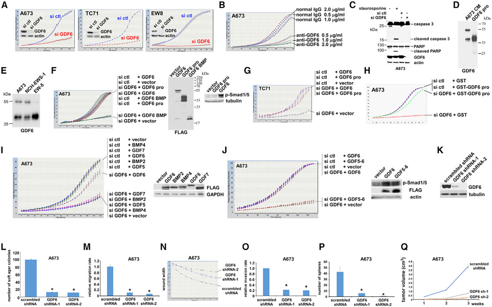
We report here that the autocrine signaling mediated by growth and differentiation factor 6 (GDF6), a member of the bone morphogenetic protein (BMP) family of cytokines, maintains Ewing sarcoma growth by preventing Src hyperactivation. Surprisingly, Ewing sarcoma depends on the prodomain, not the BMP domain, of GDF6. We demonstrate that the GDF6 prodomain is a ligand for CD99, a transmembrane protein that has been widely used as a marker of Ewing sarcoma. The binding of the GDF6 prodomain to the CD99 extracellular domain results in recruitment of CSK (C-terminal Src kinase) to the YQKKK motif in the intracellular domain of CD99, inhibiting Src activity. GDF6 silencing causes hyperactivation of Src and p21-dependent growth arrest. We demonstrate that two GDF6 prodomain mutants linked to Klippel-Feil syndrome are hyperactive in CD99-Src signaling. These results reveal a cytokine signaling pathway that regulates the CSK-Src axis and cancer cell proliferation and suggest the gain-of-function activity for disease-causing GDF6 mutants.

Keywords: CD99; CSK; Ewing sarcoma; GDF6; Klippel-Feil syndrome; Src; proteomics; secretome.


Conflict of interest statement

Declaration of Interests The authors declare no competing interests.

Figures

Figure 1.
Figure 1.. GDF6 Is a Transcriptional Target of EWS-FLI1 and Highly Expressed in Ewing Sarcoma
(A) EWS-FLI1 silencing by an shRNA targeting FLI1 C terminus results in reduced GDF6 RNA expression in Ewing sarcoma (n = 3). (B) EWS-FLI1 silencing results in reduced GDF6 protein expression in Ewing sarcoma. C, control scrambled shRNA; F, FLI1 C-terminal shRNA. (C) EWS-FLI1 induces GDF6 RNA expression in human mesenchymal stem cells, putative cells of origin of Ewing sarcoma (n = 3). (D) EWS-FLI-1 binds to the GDF6 gene promoter in Ewing sarcoma. Chromatin immunoprecipitation analysis for EWS-FLI-1 binding to the promoter of GDF6 and known EWS-FLI1 target genes (NR0B1 and EZH2), as well as control (GAPDH), with and without EWS-FLI1 silencing in A673 cells (n = 3). (E) GDF6 is highly expressed in Ewing sarcoma tumors and cell lines. GDF6 RNA expression was analyzed by quantitative real-time RT-PCR and was normalized to the levels in human mesenchymal stem cells (hMSCs). GDF6 is highly expressed in Ewing sarcoma tumors, Ewing sarcoma patient-derived xenograft tumors (NCH-EWS-1 and EW-5), and Ewing sarcoma cell lines. (n = 3)
Figure 2.
Figure 2.. Ewing Sarcoma Depends on the Prodomain of GDF6
(A) GDF6 silencing inhibits Ewing sarcoma proliferation. GDF6 was silenced by siRNA transfection in A673, TC71, and EW8 cells, and cell proliferation was assessed by IncuCyte live-cell imaging system. GDF6 silencing was verified by immunoblotting (inset). (B) Anti-GDF6 prodomain antibody inhibits Ewing sarcoma proliferation. Anti-GDF6 prodomain antibody or control normal IgG was added at the indicated concentration, and cell proliferation was assessed by IncuCyte. (C) GDF6 silencing does not induce apoptosis. GDF6 silencing in A673 cells did not result in the cleavage of caspase-3 and PARP, markers of apoptosis. Staurosporine treatment (1 μM for 4 h) serves as a positive control for apoptosis. (D) A673 cell conditioned medium expresses an approximately 55-kDa precursor and an approximately 40-kDa prodomain of GDF6. The latter comigrates with the GDF6 prodomain secreted from transfected 293T cells. The blot was probed with anti-GDF6 prodomain antibody. (E) Ewing sarcoma patient-derived xenograft tumor cells secrete GDF6. Two patient-derived xenograft tumors (NCH-EWS-1 and EW-5) were dissociated to cells, and the secretion of the GDF6 precursor and the GDF6 prodomain was assessed by anti-GDF6 immunoblotting of the conditioned media. (F) The prodomain, but not the BMP domain, of GDF6 rescues the proliferation inhibition of A673 Ewing sarcoma cells by GDF6 silencing. A673 cells were transfected with GDF6 siRNA or control siRNA and were treated with 293T cell conditioned medium expressing GDF6, GDF6 prodomain, GDF6 BMP domain (N-terminally fused to a signal peptide), or vector. Cell proliferation was assessed by IncuCyte (left). The expression of GDF6, GDF6 prodomain, and GDF6 BMP domain was verified by anti-FLAG immunoblotting (all of these are C-terminally FLAG-tagged; middle). The addition of GDF6, but not GDF6 prodomain conditioned medium, to A673 cells activated BMP signaling assessed by phosphorylated Smad1/5 (right). (G) The GDF6 prodomain rescues the proliferation inhibition of TC71 Ewing sarcoma cells by GDF6 silencing. TC71 cells were transfected with GDF6 siRNA or control siRNA and were treated with 293T cell conditioned medium expressing GDF6, GDF6 prodomain, or vector. Cell proliferation was assessed by IncuCyte. (H) GST-GDF6 prodomain rescues the proliferation inhibition by GDF6 silencing. A673 cells were transfected with GDF6 siRNA or control siRNA and were treated with GST-GDF6 prodomain or GST (500 ng/mL). Cell proliferation was assessed by IncuCyte. (I) Unlike GDF6, GDF5, GDF7, BMP2, and BMP4 are unable to rescue the proliferation inhibition by GDF6 silencing in Ewing sarcoma. A673 cells were transfected with GDF6 siRNA or control siRNA and were treated with 293T cell conditioned medium expressing GDF6, GDF5, GDF7, BMP2, BMP4, or vector. Cell proliferation was assessed by IncuCyte (left). The expression of GDF6, GDF5, GDF7, BMP2, and BMP4 was verified by anti-FLAG immunoblotting (all of these are C-terminally FLAG-tagged; right). (J) Unlike GDF6, the GDF5 prodomain-GDF6 BMP domain fusion protein (GDF5-6) is unable to rescue the proliferation inhibition by GDF6 silencing in Ewing sarcoma. A673 cells were transfected with GDF6 siRNA or control siRNA and were treated with 293T cell conditioned medium expressing GDF5-6, GDF6, or vector. Cell proliferation was assessed by IncuCyte (left). Like GDF6, GDF5-6 was able to activate BMP signaling (right). GDF5-6 and GDF6 are both C-terminally FLAG tagged. (K) shRNA-mediated silencing of GDF6 in A673 cells. A673 cells were infected with lentiviruses expressing shRNAs against GDF6 or control scrambled shRNA. After puromycin selection, GDF6 silencing was verified by immunoblotting. (L) GDF6 silencing impairs anchorage-independent growth of Ewing sarcoma. *p < 0.05 (n = 5). (M) GDF6 silencing reduces the migration rate of Ewing sarcoma assessed by Transwell assays. *p < 0.05 (n = 4). (N) GDF6 silencing reduces the migration rate of Ewing sarcoma assessed by wound closure assays. (O) GDF6 silencing reduces the invasiveness of Ewing sarcoma assessed by Matrigel invasion assays. *p < 0.05 (n = 4). (P) GDF6 silencing inhibits the tumor sphere formation of Ewing sarcoma. *p < 0.05 (n = 5). (Q) GDF6 silencing inhibits the xenograft tumorigenicity of Ewing sarcoma. *p < 0.05 (n = 5).
Figure 3.
Figure 3.. CD99 Serves as the GDF6 Prodomain Receptor in Ewing Sarcoma
(A) GDF6 prodomain co-immunoprecipitates with CD99 and CD99L2, a paralog of CD99, upon co-transfection in 293T cells. C-terminally FLAG-tagged CD99 and/or CD99L2 was co-transfected with GDF6 prodomain in 293T cells. The interaction between GDF6 prodomain and CD99/CD99L2 was assessed by anti-FLAG immunoprecipitation followed by immunoblotting for the indicated protein. (B) GDF6 prodomain interacts with GST-CD99(1–122) in vitro. GDF6 prodomain expressed in 293T cell conditioned medium was incubated with GST-CD99 extracellular domain (1–122) or GST, and the protein interaction was assessed by GST pull-down followed by immunoblotting. Asterisks denote GST-CD99(1–122) and GST. (C) GDF6 prodomain binds to Ewing sarcoma cells through CD99/CD99L2. C-terminally FLAG-tagged GDF6 prodomain secreted from transfected 293T cells was incubated for 8 h with A673 cells that were transfected with CD99 siRNA, CD99L2 siRNA, and control siRNA as indicated (“–” denotes controls without incubation). Cells were washed, and the whole-cell lysate was prepared to assess the binding of GDF6 prodomain-FLAG to the cells. GDF6 prodomain-FLAG bound to control siRNA-transfected cells, and the binding was reduced by CD99 and CD99L2 silencing. The silencing of CD99 and CD99L2 was verified by immunoblotting. (D) CD99/CD99L2 mediate the binding of GDF6 prodomain to Ewing sarcoma cells. GDF6 prodomain fused to alkaline phosphatase (AP), or AP alone was produced from transfected 293T cells and was incubated with A673 cells that were transfected with CD99 siRNA and/or CD99L2 siRNA or control siRNA as indicated. The binding of GDF6 prodomain-AP or AP to cells was visualized by AP reaction. GDF6 prodomain-AP bound to control siRNA-expressing A673 cells, and the binding was blocked by CD99 siRNA and CD99L2 siRNA. Scale bars: 20 μm. (E) GDF6 prodomain induces the internalization of CD99 expressed in HeLa cells. CD99 expressed in HeLa cells was located in the plasma membrane. GST-GDF6 prodomain caused the internalization of CD99. Scale bars: 20 μm. (F) GDF6 induces the internalization of endogenous CD99 in A673 cells. A673 cells were treated with 293T cell conditioned medium expressing vector or GDF6. The latter induced the internalization of endogenous CD99 detected by anti-CD99 immunofluorescence (left). Scale bars: 20 μm. The quantification of surface CD99 expression (right, *p < 0.05; n = 5). (G) GDF6 silencing results in accumulation of CD99 on A673 cell surface. A673 stably expressing C-terminally FLAG-tagged CD99 was transfected with control siRNA or GDF6 siRNA, and the location of CD99-FLAG was assessed by anti-FLAG immunostaining (left). Scale bars: 20 μm. GDF6 silencing caused the accumulation of CD99-FLAG on cell surface. CD99 siRNA abolished the immunostaining of CD99-FLAG. The quantification of surface CD99-FLAG expression (right, *p < 0.05; n = 5). (H) Microscale thermophoresis analysis of GDF6 prodomain-CD99 extracellular domain interaction. The interaction between C-terminally Fc-tagged CD99 extracellular domain (residues 23–122) and GDF6 prodomain (wild-type, A249E, or L289P) was analyzed by microscale thermophoresis. GDF6 prodomain mutants displayed lower KD values than wild-type GDF6 prodomain.
Figure 4.
Figure 4.. GDF6 Prodomain-CD99 Signaling Inhibits Src through Recruitment of CSK to the YQKKK Motif in the Intracellular Domain of CD99
(A) GDF6 silencing or CD99 silencing activates Src in A673 cells. Activation of Src was assessed by phosphorylation of tyrosine 419. (B) Silencing of CD99 and CD99L2 activates Src in A673 cells. (C) Inhibition of Src by GDF6 requires CD99/CD99L2 and CSK. Transfection of 293 cells with GDF6 inhibited Src (lanes 1 and 2), which was abolished by CD99 and CD99L2 silencing (lanes 3 and 4) or by CSK silencing (lanes 5 and 6). (D) Inhibition of Src by CD99 requires CSK. Transfection of 293 cells with CD99 inhibited Src (lanes 1 and 2), which was abolished by CSK silencing (lanes 3 and 4). (E) CSK co-immunoprecipitates with CD99. C-terminally HA-tagged CSK was stably expressed in A673 cells and was immunoprecipitated with anti-HA antibody. The co-precipitation of endogenous CD99 was assessed by immunoblotting. (F) Human CD99, mouse CD99, and human CD99L2 co-immunoprecipitate with CSK. C-terminally FLAG-tagged human CD99, mouse CD99, and human CD99L2 were transfected in 293T cells, and the interaction with endogenous CSK was assessed by anti-FLAG immunoprecipitation followed by anti-CSK immunoblotting. Immunoprecipitation of each FLAG-tagged protein was verified by anti-FLAG immunoblotting. (G) Conserved YQKKKLCF-like motifs in the juxtamembrane regions of human CD99, mouse CD99, and human CD99L2. (H) The YQKKK motif interacts with CSK. Human CD99 intracellular domain (148–185), human CD99 intracellular domain lacking the YQKKKLCF sequence, YQKKKLCF, or YQKKK (all N-terminally fused to GFP) was transfected in 293T cells, and the interaction with endogenous CSK was assessed by anti-GFP immunoprecipitation followed by anti-CSK immunoblotting. Immunoprecipitation of each GFP-fusion protein was verified by anti-GFP immunoblotting (bottom). (I) The deletion of the YQKKK motif abrogates CD99-CSK interaction. C-terminally FLAG-tagged CD99 or CD99 lacking the YQKKK motif was co-transfected with GFP-tagged CSK or empty GFP vector in 293T cells. The CD99-CSK interaction was assessed by anti-GFP immunoprecipitation followed by anti-FLAG immunoblotting. (J) Endogenous GDF6 stimulates CD99-CSK interaction in Ewing sarcoma. The proximity ligation assays (PLAs) were used to assess the recruitment of CSK to CD99 in A673 cells stably expressing C-terminally HA-tagged CSK. The robust PLA signals were detected between CD99 and CSK-HA, which were abolished by GDF6 silencing. Scale bars: 20 μm. The quantification of PLA signals is shown at the bottom. *p < 0.05 (n = 5). (K) GDF6 stimulates the CSK recruitment to the YQKKK motif in CD99. HeLa cells, which do not endogenously express GDF6, were transfected with CD99-FLAG and CSK-HA and then treated with 293T cell conditioned medium expressing vector or GDF6, and the interaction between CD99-FLAG and CSK-HA was assessed by the PLA (top). GDF6 conditioned medium induced the recruitment of CSK-HA to CD99 (top). The same assay was performed using CD99 lacking the YQKKK motif, which showed that GDF6 conditioned medium was unable to induce the recruitment of CSK-HA to CD99 lacking the YQKKK motif (middle). Scale bars: 20 μm. The quantification of PLA signals is shown at the bottom. *p < 0.05 (n = 5). (L) Dominant-negative Src abrogates the proliferation inhibition by GDF6 silencing in A673 cells. A673 cells stably expressing dominant-negative Src or empty vector were transfected with GDF6 siRNA or control siRNA, and cell proliferation was assessed by IncuCyte (left). Dominant-negative Src reduced the phosphorylation of Src substrates, cortactin, p130 Cas, and STAT3 (right). (M) Src mutant lacking the CSK phosphorylation site (Y530F) inhibits A673 cell proliferation. Proliferation of A673 cells stably expressing Src Y530F or empty vector was assessed by IncuCyte (left). Src Y530F increased the phosphorylation of Src substrates, cortactin, p130 Cas, and STAT3 (right). (N) CSK abrogates the proliferation inhibition by GDF6 silencing in A673 cells. A673 cells stably expressing CSK or empty vector were transfected with GDF6 siRNA or control siRNA, and cell proliferation was assessed by IncuCyte. (O) A Src inhibitor, dasatinib, abrogates the proliferation inhibition by GDF6 silencing in A673 cells. Dasatinib was used at 50 nM.
Figure 5.
Figure 5.. GDF6 Prodomain Mutants Associated with Klippel-Feil Syndrome Are Hyperactive in GDF6-CD99-Src Signaling
(A) GDF6 A249E or L289P prodomain mutant displays enhanced co-immunoprecipitation with CD99 compared with wild-type GDF6 prodomain. GDF6 prodomain, GDF6 A249E prodomain, or GDF6 L289P prodomain was co-transfected with C-terminally FLAG-tagged CD99 or FLAG vector in 293T cells as indicated, and the interaction between each GDF6 prodomain and CD99-FLAG was assessed by anti-FLAG immunoprecipitation followed by immunoblotting. (B) GDF6 A249E or L289P prodomain mutant displays enhanced interaction with CD99 in vitro. Wild-type, A249E, or L289P GDF6 prodomain expressed in 293T cell conditioned medium was incubated with C-terminally Fc-tagged CD99 extracellular domain (residues 23–122), and protein interaction was assessed by protein A Sepharose pull-down followed by immunoblotting. (C) GDF6 A249E or L289P prodomain mutant displays enhanced binding to A673 cells. Ligand-binding assays were performed as in Figure 3C. (D) GDF6 A249E or L289P prodomain mutant more strongly inhibits Src than wild-type prodomain in 293 cells. Wild-type, A249E, or L289P GDF6 prodomain was stably expressed in 293 cells, and the levels of active Src (phosphorylated on Y419) were assessed by immunoblotting. (E) GDF6 A249E or L289P mutant more strongly inhibits Src than wild-type GDF6 in A673 cells. Wild-type, A249E, or L289P GDF6 was stably expressed in A673 cells, and the levels of active phosphorylated Src were assessed by immunoblotting. (F) GDF6 A249E or L289P prodomain mutant more strongly induces CSK recruitment to CD99. The PLA was performed in HeLa cells as in Figure 4K, using 293T cell conditioned medium expressing vector, GDF6, GDF6 prodomain, GDF6 A249E prodomain, or GDF6 L289P prodomain. Scale bars: 20 μm. The quantification of PLA signals is shown at the bottom. *p < 0.05 (n = 5).

Similar articles

Cited by

References

    1. Ambros IM, Ambros PF, Strehl S, Kovar H, Gadner H, and Salzer-Kuntschik M (1991). MIC2 is a specific marker for Ewing’s sarcoma and peripheral primitive neuroectodermal tumors. Evidence for a common histogenesis of Ewing’s sarcoma and peripheral primitive neuroectodermal tumors from MIC2 expression and specific chromosome aberration. Cancer 67, 1886–1893. - PubMed
    1. Aono A, Hazama M, Notoya K, Taketomi S, Yamasaki H, Tsukuda R, Sasaki S, and Fujisawa Y (1995). Potent ectopic bone-inducing activity of bone morphogenetic protein-4/7 heterodimer. Biochem. Biophys. Res. Commun 210, 670–677. - PubMed
    1. Arbet-Engels C, Tartare-Deckert S, and Eckhart W (1999). C-terminal Src kinase associates with ligand-stimulated insulin-like growth factor-I receptor. J. Biol. Chem 274, 5422–5428. - PubMed
    1. Asai-Coakwell M, French CR, Ye M, Garcha K, Bigot K, Perera AG, Staehling-Hampton K, Mema SC, Chanda B, Mushegian A, et al. (2009). Incomplete penetrance and phenotypic variability characterize Gdf6-attributable oculo-skeletal phenotypes. Hum. Mol. Genet 18, 1110–1121. - PubMed
    1. Asai-Coakwell M, March L, Dai XH, Duval M, Lopez I, French CR, Famulski J, De Baere E, Francis PJ, Sundaresan P, et al. (2013). Contribution of growth differentiation factor 6-dependent cell survival to early-onset retinal dystrophies. Hum. Mol. Genet 22, 1432–1442. - PubMed

Publication types

MeSH terms