. 2020 Nov 4;12(21):21004-21022.
doi: 10.18632/aging.104057. Epub 2020 Nov 4.

Plasma tau predicts cerebral vulnerability in aging

Affiliations

Plasma tau predicts cerebral vulnerability in aging

Jose L Cantero et al. Aging (Albany NY). .

Abstract

Identifying cerebral vulnerability in late life may help prevent or slow the progression of aging-related chronic diseases. However, non-invasive biomarkers aimed at detecting subclinical cerebral changes in the elderly are lacking. Here, we have examined the potential of plasma total tau (t-tau) for identifying cerebral and cognitive deficits in normal elderly subjects. Patterns of cortical thickness and cortical glucose metabolism were used as outcomes of cerebral vulnerability. We found that increased plasma t-tau levels were associated with widespread reductions of cortical glucose uptake, thinning of the temporal lobe, and memory deficits. Importantly, tau-related reductions of glucose consumption in the orbitofrontal cortex emerged as a determining factor of the relationship between cortical thinning and memory loss. Together, these results support the view that plasma t-tau may serve to identify subclinical cerebral and cognitive deficits in normal aging, allowing detection of individuals at risk for developing aging-related neurodegenerative conditions.

Keywords: FDG-PET; aging; cerebral vulnerability; cortical thickness; plasma tau.


Conflict of interest statement

CONFLICTS OF INTEREST: The authors declare that they have no conflicts of interest.

Figures

Figure 1
Figure 1
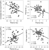
Significant correlations between plasma t-tau, plasma Aβ1-42, and memory performance. Variables included in the scatter plots correspond to the standardized residuals obtained from linear regression analyses. FCSRT: Free and Cued Selective Reminding Test; MFQ: Memory Functioning Questionnaire.
Figure 2
Figure 2
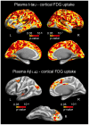
Significant associations between increased plasma t-tau/Aβ1-42 and lower cortical FDG uptake. Results are represented on inflated cortical surfaces. Left (L) and right (R). The color scale bar illustrates the range of significant p-values.
Figure 3
Figure 3
Significant associations between increased plasma t-tau/Aβ1-40/Aβ1-42 and patterns of cortical thinning. Results are represented on inflated cortical surfaces. Left (L) and right (R). The color scale bar illustrates the range of significant p-values.
Figure 4
Figure 4
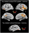
Significant associations between plasma t-tau-related reductions of cortical FDG uptake/cortical thinning and memory performance. Results are represented on inflated cortical surfaces. Left (L) and right (R). The color scale bar illustrates the range of significant p-values.
Figure 5
Figure 5
Serial mediating role of plasma t-tau and cortical FDG uptake on the relationship between cortical thinning and memory deficits in aging. Path analysis showing the serial mediation of higher plasma t-tau and lower FDG uptake in the orbitofronal cortex on the relationship between cortical thinning in the temporal lobe and memory deficits. Numbers along paths are unstandardized regression coefficients with the standard deviation in parenthesis. Asterisks indicate that the direct path as well as the total and indirect effect were statistically significant (*p < 0.05; **p < 0.005; ***p < 0.001). Thicker lines refer to the paths intervening in the three-way indirect effect.

Similar articles

Cited by

References

    1. Goedert M, Spillantini MG, Jakes R, Rutherford D, Crowther RA. Multiple isoforms of human microtubule-associated protein tau: sequences and localization in neurofibrillary tangles of Alzheimer’s disease. Neuron. 1989; 3:519–26. 10.1016/0896-6273(89)90210-9 - DOI - PubMed
    1. Binder LI, Frankfurter A, Rebhun LI. The distribution of tau in the mammalian central nervous system. J Cell Biol. 1985; 101:1371–78. 10.1083/jcb.101.4.1371 - DOI - PMC - PubMed
    1. Weingarten MD, Lockwood AH, Hwo SY, Kirschner MW. A protein factor essential for microtubule assembly. Proc Natl Acad Sci USA. 1975; 72:1858–62. 10.1073/pnas.72.5.1858 - DOI - PMC - PubMed
    1. Drubin DG, Kirschner MW. Tau protein function in living cells. J Cell Biol. 1986; 103:2739–46. 10.1083/jcb.103.6.2739 - DOI - PMC - PubMed
    1. Avila J, Lucas JJ, Perez M, Hernandez F. Role of tau protein in both physiological and pathological conditions. Physiol Rev. 2004; 84:361–84. 10.1152/physrev.00024.2003 - DOI - PubMed

Publication types

MeSH terms