Review
. 2020 Nov 2;13(11):360.
doi: 10.3390/ph13110360.

Gene Regulation Using Spherical Nucleic Acids to Treat Skin Disorders

Affiliations
Review

Gene Regulation Using Spherical Nucleic Acids to Treat Skin Disorders

Thomas R Holmes et al. Pharmaceuticals (Basel). .

Erratum in

Abstract

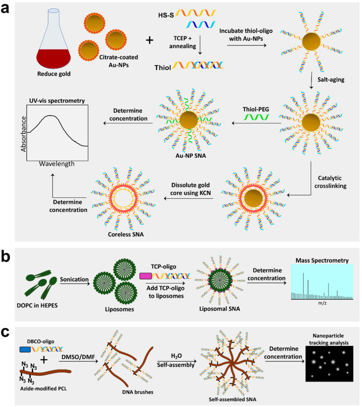
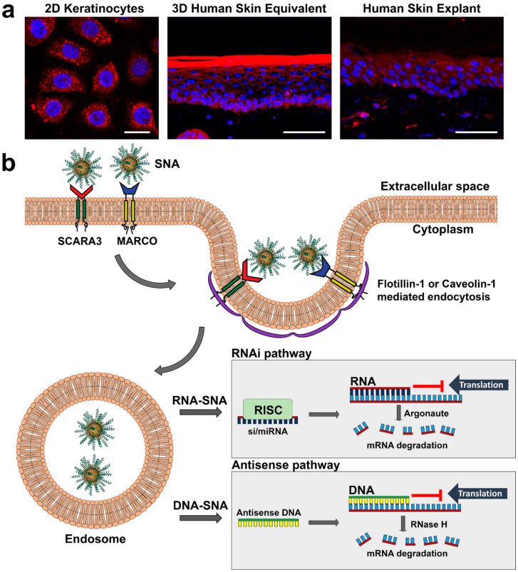
Spherical nucleic acids (SNAs) are nanostructures consisting of nucleic acids in a spherical configuration, often around a nanoparticle core. SNAs are advantageous as gene-regulating agents compared to conventional gene therapy owing to their low toxicity, enhanced stability, uptake by virtually any cell, and ability to penetrate the epidermal barrier. In this review we: (i) describe the production, structure and properties of SNAs; (ii) detail the mechanism of SNA uptake in keratinocytes, regulated by scavenger receptors; and (iii) report how SNAs have been topically applied and intralesionally injected for skin disorders. Specialized SNAs called nanoflares can be topically applied for gene-based diagnosis (scar vs. normal tissue). Topical SNAs directed against TNFα and interleukin-17A receptor reversed psoriasis-like disease in mouse models and have been tested in Phase 1 human trials. Furthermore, SNAs targeting ganglioside GM3 synthase accelerate wound healing in diabetic mouse models. Most recently, SNAs targeting toll-like receptor 9 are being used in Phase 2 human trials via intratumoral injection to induce immune responses in Merkel cell and cutaneous squamous cell carcinoma. Overall, SNAs are a valuable tool in bench-top and clinical research, and their advantageous properties, including penetration into the epidermis after topical delivery, provide new opportunities for targeted therapies.

Keywords: diabetes; gene therapy; nanoparticles; psoriasis; skin cancer; spherical nucleic acids; wound healing.


Conflict of interest statement

Paller is a consultant/advisor for Exicure Inc.

Figures

Figure 1
Figure 1
Synthesis of Au-NP core, coreless, liposomal core, and self-assembling spherical nucleic acids (SNAs). (a) Gold nanoparticle (Au-NP) SNAs are produced by reducing gold into 13 nm Au-NPs and preparing oligos by annealing sense and antisense strands and cleaving the disulfide bond to create a free thiol group. Au-NPs are incubated with thiol-oligos, salt-aged to increased oligo density, and thiol-polyethylene glycol (PEG) added to fill empty spaces. Lastly, Au-NP SNA concentration can be determined by spectrophotometry. Coreless SNAs are produced by crosslinking oligo strands on the surface of the Au-NP, then dissolving the gold core using potassium cyanide (KCN). (b) Liposomal SNAs are synthesized by sonicating 1,2-dioleoyl-sn-glycero-3-phosphocholine (DOPC) free fatty acids in HEPES buffer to create liposomes ~30 nm in diameter; liposomes are incubated with oligos containing a tocopherol (TCP) moiety for integration into liposomes. L-SNA concentration is determined by mass spectrometry. (c) Self-assembling SNAs are produced by conjugating oligos with a dibenzocyclooctone-amine (DBCO) moiety to polycaprolactone (PCL) polymers modified with azide groups in a DMSO/DMF solution to create DNA brush polymers. The DNA brushes then self-assemble in the presence of water and the concentration can be determined using nanoparticle tracking analysis.
Figure 2
Figure 2
Mechanism of SNA uptake in keratinocytes. (a) Confocal imaging of keratinocyte uptake of fluorescently (Cy5) labeled SNAs (red) in 2D, 3D and human explant culture; scale bars in (a) from left to right: 20 mm, 50 mm, 50 mm. (b) SNAs are: (i) first detected on the keratinocyte cell surface by class A scavenger receptors SCARA3 and MARCO; (ii) taken up primarily by flotillin-1-mediated endocytosis, but also caveolin-1-mediated endocytosis; (iii) deposited into endosomes; and (iv) released from endosomes to suppress gene expression via the RNAi (RNA-SNAs) or antisense (DNA-SNA) pathway; in the RNAi pathway mRNA translation is blocked by antisense RNA or target mRNA is degraded by Argonaute of the RISC complex, whereas in the antisense pathway antisense DNA can block translation or initiate mRNA degradation by RNase H.

References

    1. Mirkin C.A., Letsinger R.L., Mucic R.C., Storhoff J.J. A DNA-based method for rationally assembling nanoparticles into macroscopic materials. Nature. 1996;382:607–609. doi: 10.1038/382607a0. - DOI - PubMed
    1. Banga R.J., Chernyak N., Narayan S.P., Nguyen S.T., Mirkin C.A. Liposomal spherical nucleic acids. J. Am. Chem. Soc. 2014;136:9866–9869. doi: 10.1021/ja504845f. - DOI - PMC - PubMed
    1. Brodin J.D., Auyeung E., Mirkin C.A. DNA-mediated engineering of multicomponent enzyme crystals. Proc. Natl. Acad. Sci. USA. 2015;112:4564–4569. doi: 10.1073/pnas.1503533112. - DOI - PMC - PubMed
    1. Ruan W., Zheng M., An Y., Liu Y., Lovejoy D.B., Hao M., Zou Y., Lee A., Yang S., Lu Y., et al. DNA nanoclew templated spherical nucleic acids for siRNA delivery. Chem. Commun. 2018;54:3609–3612. doi: 10.1039/C7CC09257A. - DOI - PubMed
    1. Young K.L., Scott A.W., Hao L., Mirkin S.E., Liu G., Mirkin C.A. Hollow spherical nucleic acids for intracellular gene regulation based upon biocompatible silica shells. Nano Lett. 2012;12:3867–3871. doi: 10.1021/nl3020846. - DOI - PMC - PubMed