. 2020 Nov 2;9(11):1590.
doi: 10.3390/foods9111590.

Unripe Black Raspberry (Rubus coreanus Miquel) Extract and Its Constitute, Ellagic Acid Induces T Cell Activation and Antitumor Immunity by Blocking PD-1/PD-L1 Interaction

Affiliations

Unripe Black Raspberry (Rubus coreanus Miquel) Extract and Its Constitute, Ellagic Acid Induces T Cell Activation and Antitumor Immunity by Blocking PD-1/PD-L1 Interaction

Ji Hye Kim et al. Foods. .

Abstract

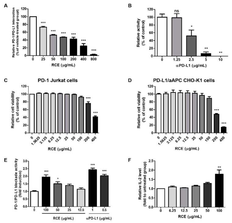
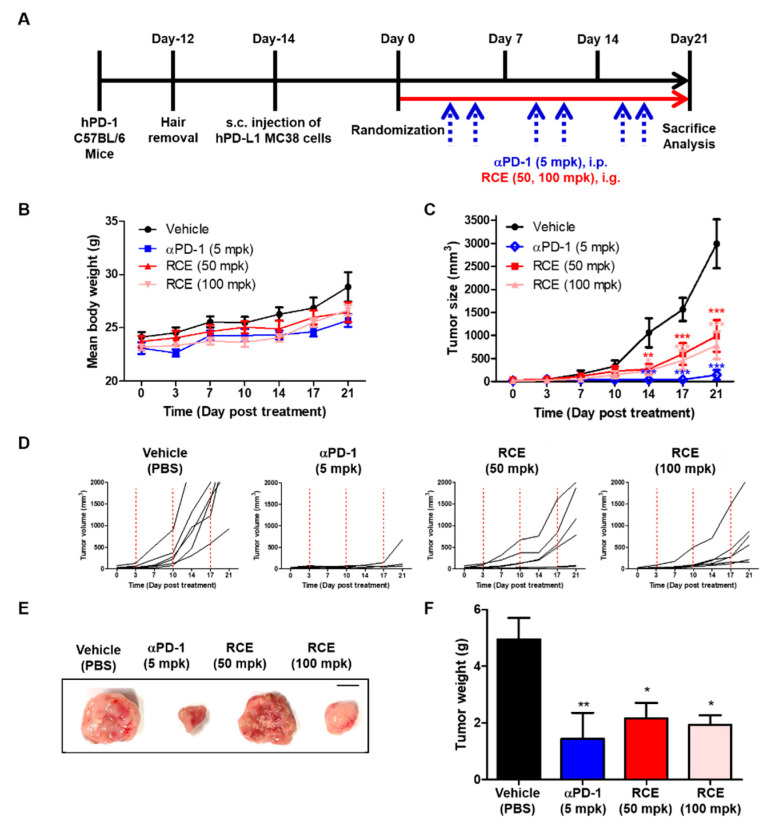
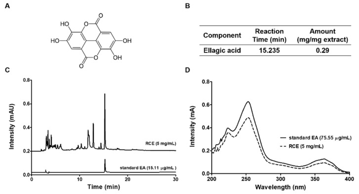
Rubus coreanus Miquel (R. coreanus) is a unripen fruit of black raspberry native to eastern Asia. It is used as traditional oriental medicine and supplementary foods for centuries. Previous studies have shown that the R. coreanus extract (RCE) and its main constitute ellagic acid possess diverse biological activities. However, the effects of RCE on antitumor immunity and T cell function were not fully understood. The present study describes the anti-tumor effect of RCE in humanized PD-1 mice by blocking PD-1/PD-L1 interaction. Competitive enzyme-linked immunosorbent assay (ELISA) and pull down assay were performed to elucidate the binding properties of RCE in vitro. Cellular PD-1/PD-L1 blockade activities were measured by T cell receptor (TCR)-induced nuclear factor of activated T cells-luciferase activity in co-cultured cell models with PD-1/NFAT Jurkat and PD-L1/aAPC CHO-K1 cells. The in vivo efficacy of RCE was confirmed in humanized PD-1 mice bearing MC38 colorectal tumor. RCE and ellagic acid dose-dependently block the binding of PD-1 to PD-L1. Moreover, oral administration of RCE showed the potent anti-tumor activity similar to anti-PD-1 antibody. The present study suggests that RCE possesses potent anti-tumor effect via PD-1/PD-L1 blockade, and ellagic acid is the main compound in RCE. Thus, we provide new aspects of RCE as an immunotherapeutic agent.

Keywords: T cell function; antitumor immunity; black raspberry; ellagic acid; programmed cell death protein 1; programmed death-ligand 1.


Conflict of interest statement

The authors declare no conflict of interest.

Figures

Figure 1
Figure 1
Effect of Rubus coreanus extract (RCE) on programmed cell death protein 1 (PD-1)/PD-1 ligand-1 (PD-L1) protein interaction using competitive enzyme-linked immunosorbent assay (ELISA) or cell-based assay. (A,B) The effect of RCE on the binding of PD-1 and PD-L1 in vitro using a competitive ELISA assay. (C,D) The effect of RCE on cell viability of PD-1/NFAT Jurkat cells (C) and PD-L1/aAPC CHO-K1 cells (E) using the Cell Counting Kit-8 (CCK) assay. Each of the cells were treated according to the indicated concentrations of RCE for 24 h. (E) The effect of RCE on cellular PD-1/PD-L1 blockade activity. PD-1/NFAT Jurkat effector cells and PD-L1/aAPC CHO-K1 target cells were co-cultured with RCE or αPD-L1 for 24 h at the indicated concentrations. The relative PD-1/PD-L1 blockade activity were measured by PD-1/NFAT luciferase reporter assay. (F) Effect of RCE on IL-2 cytokine release in co-cultured cell model. Cell culture media was analyzed to measure IL-2 level by cytokine ELISA. Data are presented as mean ± S.E. of three representative independent experiments. Asterisks indicate significant inhibition of PD-1/PD-L1 binding activity by each test inhibitor as compared with the vehicle-treated group; *** p < 0.001, ** p < 0.01, * p < 0.05, compared with the vehicle group.
Figure 2
Figure 2
Anti-tumor efficacy of RCE on hPD-L1 MC38 tumor in humanized PD-1 mice. (A) Experimental scheme. (B) Mean bodyweight. (C) Mean tumor growth curves. (D) The individual tumor growth over time for plot. MC38 cells expressing hPD-L1 (1.5 × 10⁶ cells/mL) were injected subcutaneously (day-14) into humanized PD-1 mice (3 × 10⁵ cells/200 μL in PBS/mouse). Once tumors became palpable (~100 mm3), the tumor-bearing mice were randomly assigned to either a vehicle group or treatment group (n = 6). Animals were treated with vehicle (PBS, 10 mL/kg, q.d., i.g.), αPD-1 (5 mpk, b.i.w., i.p.), RCE (50 mpk, q.d., i.g.) and RCE (100 mpk, q.d., i.g.). Tumor mice received an equivalent volume of vehicle (200 μL). On day 21 post administration, all mice were sacrificed for in vivo analysis. (E) Representative images of subcutaneous tumor mass on day 21 after treatment (scale bars, 1 cm). (F) Mean tumor weight. Statistics were analyzed using two-way ANOVA with Bonferroni post-hoc test; *** p < 0.001, ** p < 0.01, * p < 0.05, compared with the vehicle group.
Figure 3
Figure 3
Identification of the main compound of RCE by HPLC analysis. (A) Structures of the major compound of RCE. (B) The amount of ellagic acid in RCE and retention time. (C) High-performance liquid chromatography (HPLC) profiles of ellagic acid from RCE. (D) Standard ellagic acid was used for reference compound and monitored at 254 nm.
Figure 4
Figure 4
Effect of ellagic acid on PD-1/PD-L1 interaction in vitro. (A) Effect of ellagic acid on PD-1/PD-L1 interaction was measured by competitive ELISA. Ellagic acid was treated as indicated concentration (0–120.9 μg/mL). (B) Ellagic acid binds to PD-1 and PD-L1. Pull-down assays were performed to detect the binding of ellagic acid with PD-1 or PD-L1. Binding by ellagic acid between human recombinant PD-1 or PD-L1 proteins were confirmed by Western blot using antibodies against PD-1 (upper panel) or PD-L1 (lower panel). Lane 1: Recombinant PD-1 or PD-L1 protein alone; lane 2: each of the proteins were precipitated with Sepharose 4B-alone; lane 3: each of the proteins were precipitated with ellagic acid–Sepharose 4B beads (EA-Sepharose 4B). (C,D) Effect of ellagic acid on cell viability of (c) PD-1/NFAT Jurkat cells or (D) PD-L1/aAPC PD-L1 CHO-K1 cells. The cell viabilities were analyzed using the Cell Counting Kit-8 (CCK) assay. Cells were treated with or ellagic acid as indicated concentrations (0–120.9 μg/mL). (E) Effect of ellagic acid on cellular PD-1/PD-L1 blockade activity. PD-1/NFAT Jurkat effector cells and PD-L1/aAPC CHO-K1 target cells were co-cultured with ellagic acid for 48 h at the indicated concentrations (0–7.56 μg/mL). The relative PD-1/PD-L1 blockade activity were measured by PD-1/NFAT luciferase reporter assay. (F) Effect of ellagic acid on IL-2 cytokine release in co-cultured cell model. Cell culture media was analyzed to measure IL-2 level by cytokine ELISA. Data are presented as mean ± S.E. of three representative independent experiments. Asterisks indicate significant inhibition of PD-1/PD-L1 binding activity by each test inhibitor as compared with the vehicle-treated group; *** p < 0.001, ** p < 0.01, * p < 0.05, compared with the vehicle group.

References

    1. Kaufman H.L., Atkins M.B., Subedi P., Wu J., Chambers J., Joseph Mattingly T., II, Campbell J.D., Allen J., Ferris A.E., Schilsky R.L., et al. The promise of Immuno-oncology: Implications for defining the value of cancer treatment. J. Immunother. Cancer. 2019;7:129. doi: 10.1186/s40425-019-0594-0. - DOI - PMC - PubMed
    1. Skalniak L., Zak K.M., Guzik K., Magiera K., Musielak B., Pachota M., Szelazek B., Kocik J., Grudnik P., Tomala M., et al. Small-molecule inhibitors of PD-1/PD-L1 immune checkpoint alleviate the PD-L1-induced exhaustion of T-cells. Oncotarget. 2017;8:72167–72181. doi: 10.18632/oncotarget.20050. - DOI - PMC - PubMed
    1. Calik I., Calik M., Turken G., Ozercan I.H., Dagli A.F., Artas G., Sarikaya B. Intratumoral Cytotoxic T-Lymphocyte Density and PD-L1 Expression Are Prognostic Biomarkers for Patients with Colorectal Cancer. Medicina. 2019;55:723. doi: 10.3390/medicina55110723. - DOI - PMC - PubMed
    1. Ganesan A., Ahmed M., Okoye I., Arutyunova E., Babu D., Turnbull W.L., Kundu J.K., Shields J., Agopsowicz K.C., Xu L., et al. Comprehensive in vitro characterization of PD-L1 small molecule inhibitors. Sci. Rep. 2019;9:12392. doi: 10.1038/s41598-019-48826-6. - DOI - PMC - PubMed
    1. Akinleye A., Rasool Z. Immune checkpoint inhibitors of PD-L1 as cancer therapeutics. J. Hematol. Oncol. 2019;12:92. doi: 10.1186/s13045-019-0779-5. - DOI - PMC - PubMed