Review
. 2020 Nov 2;9(11):2401.
doi: 10.3390/cells9112401.

The Microbiota and Gut-Related Disorders: Insights from Animal Models

Affiliations
Review

The Microbiota and Gut-Related Disorders: Insights from Animal Models

Layla Kamareddine et al. Cells. .

Abstract

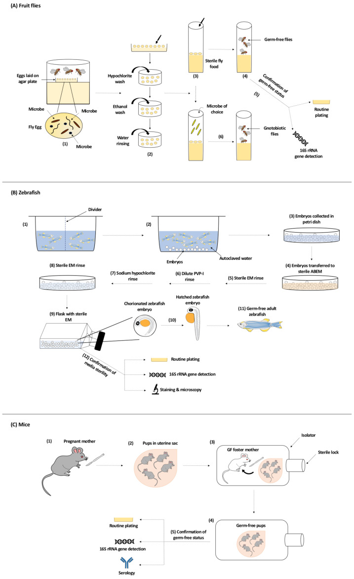
Over the past decade, the scientific committee has called for broadening our horizons in understanding host-microbe interactions and infectious disease progression. Owing to the fact that the human gut harbors trillions of microbes that exhibit various roles including the production of vitamins, absorption of nutrients, pathogen displacement, and development of the host immune system, particular attention has been given to the use of germ-free (GF) animal models in unraveling the effect of the gut microbiota on the physiology and pathophysiology of the host. In this review, we discuss common methods used to generate GF fruit fly, zebrafish, and mice model systems and highlight the use of these GF model organisms in addressing the role of gut-microbiota in gut-related disorders (metabolic diseases, inflammatory bowel disease, and cancer), and in activating host defense mechanisms and amending pathogenic virulence.

Keywords: animal models; germ-free; gut microbiota; gut-related disorders; host–defense; pathogen virulence.


Conflict of interest statement

The authors declare no conflict of interest.

Figures

Figure 1
Figure 1
The gut structure and normal flora of different model organisms including (AC) the fruit fly, (DF) zebrafish, and (GI) mouse. (A,D,G) Anatomy of the gut; (B,E,H) cell types present, including enterocytes (EC), enteroendocrine cells (EE), intestinal stem cells (ISC), enteroblasts (EB), goblet cells, stem cells (SC), vacuolated cells (VC) and, in the mouse, chemosensory tuft cells in the villi and paneth cells, transit amplifying cells (TAC), and quiescent ISC in the crypts; (C,F,I) composition of the major phyla according to [59,60,61]. BM = basement membrane.
Figure 2
Figure 2
Generation of Germ-Free Model Organisms. (A) Generation of Germ-Free Fruit Flies. Flies trapped in a cage lay their eggs on the agar plate. The surface of the laid eggs harbors microorganisms from maternal fecal deposits (1). Using a sterile paint brush, eggs are collected from the agar plate before larval formation, dechorionated with hypochlorite solution, and rinsed with water. An ethanol wash between the hypochlorite dechorionation step and the water rinsing step could be done (2). Dechorionated eggs are transferred to sterile fly food (3) and germ-free flies are generated (4). The most common methods used to confirm the germ-free status of flies include routine culturing on selective media permissive to the growth of gut bacteria and 16S rRNA gene detection via PCR amplification and sequencing (5). To generate gnotobiotic flies, microbe of choice is added to the sterile fly food vial containing dechorionated eggs (6). (B) Generation of Germ-Free Zebrafish. Adult zebrafish are set up in breeding cages overnight with dividers to prevent breeding and spawning (1). The next day, adults are transferred to tanks with autoclaved water and dividers are removed to allow breeding and spawning for less than an hour. Embryos are deposited in the tanks (2) and are then collected into petri dishes and rinsed thoroughly to remove any debris (3). In a biosafety cabinet, zebrafish embryos are transferred to petri dishes with sterile antibiotic embryo media (ABEM) (4) and undergo a sterile embryo media (EM) rinse (3 times) (5), followed by a dilute polyvinylpyrrolidone-iodine (PVP-I) rinse (6), and finally a sodium hypochlorite rinse (7). Embryos are then rinsed with sterile EM (3 times) again (8) before being transferred into sterile tissue culture flasks containing sterile EM where they will be raised (9). If hatching (10) has not occurred within 3 days post-fertilization, gentle flicking of the flask can mechanically dechorionated the embryos. Embryos eventually develop into germ-free adult zebrafish (11). Commonly used methods to test the sterility of the zebrafish media are routine culturing on various selective media under aerobic and anaerobic conditions, 16S rRNA gene detection via PCR amplification and sequencing, and staining and microscopy procedures such as fluorescence in situ hybridization and Gram stain (12). (C) Generation of Germ-Free Mice. Pups are removed from their mother by Caesarean section (1) and while still in the uterine sac (2) transferred to a germ-free foster mother (also raised germ-free) in an isolator (3). The first generation is not used for experiments, since the mother was not germ-free and may have transmitted microbes transplacentally to the fetus. Further generations of pups are delivered and cleaned inside this isolator for experimental use (4). The germ-free status of the animals is monitored by routine plating of stool material, detection of 16S rRNA by PCR amplification and sequencing, and serological techniques (5).

References

    1. Dominguez-Bello M.G., Blaser M.J., Ley R.E., Knight R. Development of the human gastrointestinal microbiota and insights from high-throughput sequencing. Gastroenterology. 2011;140:1713–1719. doi: 10.1053/j.gastro.2011.02.011. - DOI - PMC - PubMed
    1. O’Hara A.M., Shanahan F. The gut flora as a forgotten organ. EMBO Rep. 2006;7:688–693. doi: 10.1038/sj.embor.7400731. - DOI - PMC - PubMed
    1. Wang B., Yao M., Lv L., Ling Z., Li L. The Human Microbiota in Health and Disease. Engineering. 2017;3:71–82. doi: 10.1016/J.ENG.2017.01.008. - DOI
    1. Fritz J.V., Desai M.S., Shah P., Schneider J.G., Wilmes P. From meta-omics to causality: Experimental models for human microbiome research. Microbiome. 2013;1:14. doi: 10.1186/2049-2618-1-14. - DOI - PMC - PubMed
    1. Le Chatelier E., Nielsen T., Qin J., Prifti E., Hildebrand F., Falony G., Almeida M., Arumugam M., Batto J.M., Kennedy S., et al. Richness of human gut microbiome correlates with metabolic markers. Nature. 2013;500:541–546. doi: 10.1038/nature12506. - DOI - PubMed

Publication types

LinkOut - more resources