. 2020 Nov 2;12(11):1250.
doi: 10.3390/v12111250.

Functional Analyses of Bovine Foamy Virus-Encoded miRNAs Reveal the Importance of a Defined miRNA for Virus Replication and Host-Virus Interaction

Affiliations

Functional Analyses of Bovine Foamy Virus-Encoded miRNAs Reveal the Importance of a Defined miRNA for Virus Replication and Host-Virus Interaction

Wenhu Cao et al. Viruses. .

Abstract

In addition to regulatory or accessory proteins, some complex retroviruses gain a repertoire of micro-RNAs (miRNAs) to regulate and control virus-host interactions for efficient replication and spread. In particular, bovine and simian foamy viruses (BFV and SFV) have recently been shown to express a diverse set of RNA polymerase III-directed miRNAs, some with a unique primary miRNA double-hairpin, dumbbell-shaped structure not known in other viruses or organisms. While the mechanisms of expression and structural requirements have been studied, the functional importance of these miRNAs is still far from understood. Here, we describe the in silico identification of BFV miRNA targets and the subsequent experimental validation of bovine Ankyrin Repeat Domain 17 (ANKRD17) and Bax-interacting factor 1 (Bif1) target genes in vitro and, finally, the suppression of ANKRD17 downstream genes in the affected pathway. Deletion of the entire miRNA cassette in the non-coding part of the U3 region of the long terminal repeats attenuated replication of corresponding BFV mutants in bovine cells. This repression can be almost completely trans-complemented by the most abundant miRNA BF2-5p having the best scores for predicted and validated BFV miRNA target genes. Deletion of the miRNA cassette does not grossly affect particle release and overall particle composition.

Keywords: ANKRD17; Bif1 (SH3GLB1); bovine foamy virus; innate immunity; miRNA expression; miRNA target gene identification; replication in vitro; spumaretrovirus; virus-host-interaction.


Conflict of interest statement

The authors declare no conflict of interest.

Figures

Figure 1
Figure 1
Deletion of the miRNA cassette results in reduced infectivity in bovine MICL reporter cells. Bar diagram of the virus titers (FFU/mL) expressed as number of GFP-positive focus forming units (FFU) from three independent experiments. Low-density MICL cells were infected with serially diluted wt virus (Riem 34 and MDBK 24) and the corresponding miRNA cassette deletion mutants (Riems 34-ΔmiRNA and MDBK 24-ΔmiRNA) originating from transfected HEK293T cells. Five days p.i., viral titers were determined by fluorescence microscopy of GFP-positive cell foci due to the BFV infection-mediated induction of GFP expression. Error bars are given and differences between titers of the wt and the ΔmiRNA mutant virus were analyzed by the Welch’s t-test and found to be highly significant (***, p < 0.001).
Figure 2
Figure 2
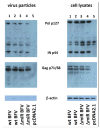
Deletion of the miRNA cassette neither affects BFV Gag and Pol expression and processing nor BFV particle release and composition in transfected and MDBK-co-cultured HEK293T cells. Each two sub-clones of plasmids encoding high titer pCMV-BFV-MDBK 24 (wt BFV) and pCMV-BFV-MDBK 24-ΔmiRNA (ΔmiR BFV) and the pcDNA2.1 control plasmid were transfected into HEK293T cells together with an EGFP expression plasmid using the PEI method. Two days later, MDBK cells were added and three days later, cells were harvested for protein analyses. Cell culture supernatants were used to enrich BFV particles by sucrose cushion sedimentation. Each 15 µg of cell lysates and regular aliquots of enriched particles were subjected to immunoblotting (as given above the panels) using a cross-reactive rabbit anti PFV IN antiserum [39] (top panels) and a serum pool from BFV-infected cattle (middle panels). The BFV-specific proteins detected are labeled between the panels. A directly conjugated antibody against β-actin served as the loading control (bottom panels).
Figure 3
Figure 3
Trans-complementation of miRNA-deficient BFV with miR-BF2-5p restores BFV titers in bovine MICL cells. Bar diagrams of the virus titers (FFU/mL) and the number of GFP-positive focus forming units from three independent experiments. (A) MICL cells were infected first with serially diluted miRNA cassette deletion mutants (BFV-Riems 34-ΔmiRNA and BFV-MDBK 24-ΔmiRNA). Six h after infection, the corresponding miR-BF2-5p mimic and negative control were transfected. (B) MICL cells were transfected first with the miR-BF2-5p mimic and negative control. Six h after transfection, the cell were infected with serially diluted miRNA cassette deletion mutants (BFV-Riems 34-ΔmiRNA and BFV-MDBK 24-ΔmiRNA). At 4 day p.i., viral titers were determined in both experiments by fluorescence microscopy. Error bars for the titers determined are given and differences between titers of the viruses trans-complemented with the BF2-5p mimic and control miRNA were analyzed by the Welch’s t-test (***, p < 0.001).
Figure 4
Figure 4
MiR-BF2-5p binding site prediction within ANKRD17 (panel A) and Bif1 (panel B). The overall bovine ANKRD17 and Bif1 genes consist of 5’UTR, the open reading frame (ORF), and the 3′UTR. Based on sequence complementarity analysis, two possible binding sites (a and b) were identified in the 3′UTR of ANKRD17 starting 85 and 448 nt downstream of the stop codon (A), the sequences which are complementary to the seed sequence of miR-BF2-5p, are shown in red. For Bif1 (B), three possible binding sites (a-b-a) were found in the 3′UTR, starting 217, 245, and 284 nt downstream of the stop codon, the sequences which are complementary to the seed sequence of miR-BF2-5p are shown in red.
Figure 5
Figure 5
Binding efficacy of miR-BF2-5p and ANKRD17 (A) and Bif1 (B) target sequences in BFV-infected cells. MDBK and BoMac cells (as given) were infected with high-titer BFV-MDBK24 (wt BFV) and miRNA-deficient BFV-MDBK24 (ΔmiRNA) at a MOI of 0.1 or cell culture medium was added (MOCK). Two days p.i., the reporter plasmids psiCHECK2-ANKRD17-original (A.Ori, panel A) and psiCHECK2-Bif1-original (B.Ori, panel B) were transfected into the cells. At 24 h post-transfection, cell lysates were harvested and analyzed for the suppression of Rluc activity in standard DLR assays as described. Normalized luciferase data (Renilla versus firefly luciferase activity) are presented as bar diagrams of three independent experiments. Differences between treated and control groups (MOCK) were analyzed by the Welch’s t-test (***, p < 0.001).
Figure 6
Figure 6
Binding efficacy of miR-BF2-5p mimic and the target sequences in the 3′UTR of bovine ANKRD17 (A) and Bif1 (B). HEK293T, MDBK, and BoMac cells were transfected with four reporter plasmid/RNA oligonucleotide combinations (letters A and B refer to the ANKRD17 and Bif1 target genes and suffixes Ori and Scr refer to original (wild-type) and scrambled (non-functional control) target sequences, respectively): A.Scr/B.Scr plus miR-BF2-5p mimic (A.Scr/B.Scr (2–5p mimic)); A.Ori/B.Ori plus miR-BF2-5p mimic (A.Ori/B.Ori (2–5p mimic)); A.Scr/B.Scr plus mimic NC (A.Scr/B.Scr (mimic NC)); A.Ori/B.Ori plus mimic NC (A.Ori/B.Ori (mimic NC)). In addition, the empty DLR report plasmid was used as control (black bars). At 24 h post transfection, cell lysates were harvested and analyzed for the suppression of Rluc activity in standard DLR assays as described. Normalized luciferase data (Renilla versus firefly luciferase activity) are presented as bar diagrams of three independent experiments. Differences between treated groups and MOCK or Control groups were analyzed by the Welch’s t-test (***, p < 0.001; **, p < 0.01).
Figure 7
Figure 7
Validation of ANKRD17 (A) and Bif1 (B) mRNAs as direct targets of miR-BF2-5p. MDBK cells were transfected with miR-BF2-5p mimic and mimic NC (negative control), or U6 promoter driven cassette and U6 promoter empty plasmid (NC), or infected with wt BFV-MDBK24 and ΔmiRNA BFV-MDBK24. After three days, transfected, infected, and untreated cells (MOCK) were harvested and RNA was extracted for qRT-PCR analysis. Expression data normalized relative to GAPDH RNA are shown as bar diagrams of three independent experiments. Differences between treated groups and MOCK or negative control groups were analyzed by the Welch’s t-test (*, p < 0.05).
Figure 8
Figure 8
Validation of steady-state protein levels of ANKRD17 (A) and Bif1 (B) as direct targets of miR-BF2-5p. MDBK cells were transfected with miR-BF2-5p mimic and mimic NC (negative control), or U6 promoter driven cassette and U6 promoter empty plasmid (NC), or infected with wt BFV-MDBK24 and ΔmiRNA BFV-MDBK24. After three days, the transfected and infected cells were harvested and protein was extracted for western blotting. The expression of β-actin was monitored as a control for proper protein loading. In this blot representing one out of two experiments, the 75 kDa splice variant of bovine ANKRD17 and the full-length 40 kDa Bif1 proteins were specifically detected using antibodies as described in Section 2.7.
Figure 9
Figure 9
Suppression of pro-inflammatory signaling via IFN-β (A) and NF-κB by (B) miR-BF2-5p. BoMac cells were infected with wt BFV-MDBK24 and 2 d p.i. transfected with miR-BF2-5p inhibitor and inhibitor negative control (NC) RNA oligonucleotides. In parallel, another group of BoMac cells were infected with ΔmiRNA BFV-MDBK24 and 2 d p.i. transfected with miR-BF2-5p mimic and mimic NC RNA oligonucleotides. At 48 h after transfection, cells were harvested and total RNA was extracted for qRT-PCR analyses, testing the expression level of IFN-β and NF-κB, respectively. Expression data normalized relative to GAPDH RNA are shown as bar diagrams of three independent experiments. For comparison, the relative and GAPDH-normalized expression levels of IFN-β and NF-κB of untreated BoMac cells are given (MOCK). Differences between treated groups and MOCK or negative control groups were analyzed by the Welch’s T-test (*, p < 0.05).
Figure 10
Figure 10
Kinetics of ANKRD17 and Bif1 steady state levels in BFV-infected and mock-infected BoMac (lanes 1 to 8) and MDBK cells (lanes 9 to 16). Sub-confluent BoMac and MDBK cells were either mock-infected (lanes 1 to 4 and 9 to 12) or infected with wt BFV-MDBK24 at a MOI of approximately 0.1 (lanes 5 to 8 and 13 to 16) and harvested 1, 2, 3, and 4 p.i. (as indicated below the blots). Identical amounts of protein were loaded and β-actin expression was monitored for proper protein loading (bottom panel). In this blot, the 75kDa splice variant of bovine ANKRD17 (top panel) was detected using the antiserum provided by T. Kufer and the full-length 40 kDa Bif1 protein was detected using the commercially available antiserum (middle panel). The bands specific for the 75 kDa ANKRD17 form and the 40 kDa Bif and 42 kDa β-actin are shown. Below the blots, the steady state protein levels were determined by densitometry, normalized to β-actin levels, and expression levels are displayed relative to the 1 d mock- and BFV-infected BoMac and MDBK cell values.

Similar articles

Cited by

References

    1. Cullen B.R. MicroRNA expression by an oncogenic retrovirus. Proc. Natl. Acad. Sci. USA. 2012;109:2695–2696. doi: 10.1073/pnas.1200328109. - DOI - PMC - PubMed
    1. Cullen B.R. MicroRNAs as mediators of viral evasion of the immune system. Nat. Immunol. 2013;14:205–210. doi: 10.1038/ni.2537. - DOI - PMC - PubMed
    1. Bartel D.P. Metazoan MicroRNAs. Cell. 2018;173:20–51. doi: 10.1016/j.cell.2018.03.006. - DOI - PMC - PubMed
    1. Skalsky R.L., Cullen B.R. Viruses, microRNAs, and host interactions. Annu. Rev. Microbiol. 2010;64:123–141. doi: 10.1146/annurev.micro.112408.134243. - DOI - PMC - PubMed
    1. Yao Y., Smith L.P., Nair V., Watson M. An avian retrovirus uses canonical expression and processing mechanisms to generate viral microRNA. J. Virol. 2014;88:2–9. doi: 10.1128/JVI.02921-13. - DOI - PMC - PubMed

Publication types