

Rescue Potential of Supportive Embryo Culture Conditions on Bovine Embryos Derived from Metabolically Compromised Oocytes
- PMID: 33147848
- PMCID: PMC7663530
- DOI: 10.3390/ijms21218206
Rescue Potential of Supportive Embryo Culture Conditions on Bovine Embryos Derived from Metabolically Compromised Oocytes
Abstract
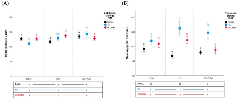
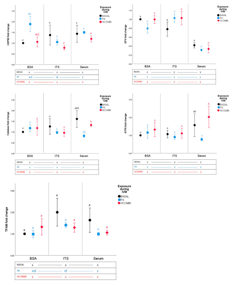
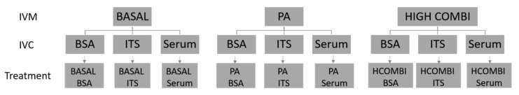
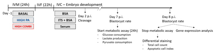
Elevated non-esterified fatty acid (NEFA), predominantly palmitic acid (PA), concentrations in blood and follicular fluid are a common feature in maternal metabolic disorders such as obesity. This has a direct negative impact on oocyte developmental competence and the resulting blastocyst quality. We use NEFA-exposure during bovine oocyte in vitro maturation (IVM) as a model to mimic oocyte maturation under maternal metabolic stress conditions. However, the impact of supportive embryo culture conditions on these metabolically compromised zygotes are not known yet. We investigated if the addition of anti-apoptotic, antioxidative and mitogenic factors (namely, Insulin-Transferrin-Selenium (ITS) or serum) to embryo culture media would rescue development and important embryo quality parameters (cell proliferation, apoptosis, cellular metabolism and gene expression patterns) of bovine embryos derived from high PA- or high NEFA-exposed oocytes when compared to controls (exposed to basal NEFA concentrations). ITS supplementation during in vitro culture of PA-exposed oocytes supported the development of lower quality embryos during earlier development. However, surviving blastocysts were of inferior quality. In contrast, addition of serum to the culture medium did not improve developmental competence of PA-exposed oocytes. Furthermore, surviving embryos displayed higher apoptotic cell indices and an aberrant cellular metabolism. We conclude that some supportive embryo culture supplements like ITS and serum may increase IVF success rates of metabolically compromised oocytes but this may increase the risk of reduced embryo quality and may thus have other long-term consequences.
Keywords: fertility; free fatty acids; in vitro culture supplementation.
Conflict of interest statement
The authors declare no conflict of interest.
Figures

References
-
- WHO . Low Fertility-the Future of Europe? (Entre Nous) WHO; Geneva, Switzerland: 2006.
-
- Jungheim E.S., Louden E.D., Chi M.M., Frolova A.I., Riley J.K., Moley K.H. Preimplantation exposure of mouse embryos to palmitic acid results in fetal growth restriction followed by catch-up growth in the offspring. Biol. Reprod. 2011;85:678–683. doi: 10.1095/biolreprod.111.092148. - DOI - PMC - PubMed
-
- Leroy J.L., Vanholder T., Mateusen B., Christophe A., Opsomer G., de Kruif A., Genicot G., Van Soom A. Non-esterified fatty acids in follicular fluid of dairy cows and their effect on developmental capacity of bovine oocytes in vitro. Reproduction. 2005;130:485–495. doi: 10.1530/rep.1.00735. - DOI - PubMed
-
- Robker R.L., Akison L.K., Bennett B.D., Thrupp P.N., Chura L.R., Russell D.L., Lane M., Norman R.J. Obese women exhibit differences in ovarian metabolites, hormones, and gene expression compared with moderate-weight women. J. Clin. Endocrinol. Metab. 2009;94:1533–1540. doi: 10.1210/jc.2008-2648. - DOI - PubMed
MeSH terms
Substances
Grants and funding
LinkOut - more resources
Full Text Sources
