. 2020 Nov 2;9(11):3542.
doi: 10.3390/jcm9113542.

Inhibition of Human Neutrophil Functions In Vitro by Multiple Sclerosis Disease-Modifying Therapies

Affiliations

Inhibition of Human Neutrophil Functions In Vitro by Multiple Sclerosis Disease-Modifying Therapies

Sara Scutera et al. J Clin Med. .

Abstract

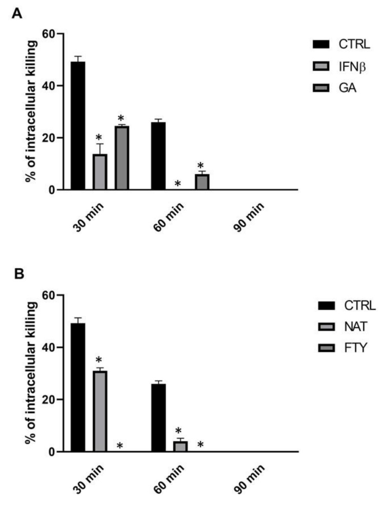
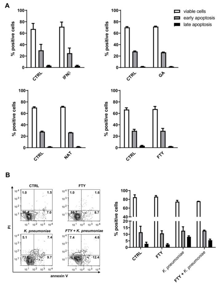
There is a growing optimism about the potential of new disease-modifying therapies (DMTs) in the management of relapsing-remitting multiple sclerosis (RRMS) patients. However, this initial enthusiasm has been tempered by evidence indicating that multiple sclerosis (MS) patients undergoing DMT may be at higher risk of developing infections through incompletely understood mechanisms. As neutrophils provide the first line of defense against pathogens, here we have compared the effects of some of the commonly used MS DMTs (i.e., moderate-efficacy injective, first-line: interferonβ-1b (IFNβ-1b), glatiramer acetate (GA); and high-efficacy, second-line: fingolimod (FTY) and natalizumab (NAT)) on the in vitro viability and functions of neutrophils isolated from healthy subjects. All the DMTs tested impaired the ability of neutrophils to kill Klebsiella pneumoniae, whereas none of them affected the rate of neutrophil apoptosis or CD11b and CD62L cell surface expression. Intriguingly, only FTY exposure negatively affected K. pneumoniae-induced production of reactive oxygen species (ROS) in polymorphonuclear leukocytes (PMNs). Furthermore, neutrophils exposed to K. pneumoniae secreted enhanced amounts of CXCL8, IL-1β and TNF-α, which were differentially regulated following DMT pretreatment. Altogether, these findings suggest that DMTs may increase the susceptibility of MS patients to microbial infections, in part, through inhibition of neutrophil functions. In light of these data, we recommend that the design of personalized therapies for RRMS patients should take into account not just the mechanism of action of the chosen DMT but also the potential risk of infection associated with the administration of such therapeutic compounds to this highly vulnerable population.

Keywords: Klebsiella pneumoniae; disease-modifying therapies; multiple sclerosis; neutrophil functions.


Conflict of interest statement

The authors declare no conflict of interest.

Figures

Figure 1
Figure 1
DMT treatment determined a reduction of intracellular K. pneumoniae killing. PMNs (106 cells/mL) were incubated for 60 min with the specific DMT (A, first-line and B, second-line), then bacteria (107 CFU/mL) were added, and their killing activity was evaluated at T30, T60 and T90. Data are shown as killing percentages from at least three independent experiments. * p < 0.01 vs. DMTs-treated cells by using the Mann–Whitney test for nonparametric data.
Figure 2
Figure 2
DMT treatment does not promote PMN apoptosis. Cell apoptosis was measured by annexin V-fluorescein isothiocyanate (FITC) and propidium iodide (PI) staining. (A) Human neutrophils were treated with the indicated DMT (IFNβ-1b, GA, NAT or FTY) for 3 h and then analyzed by flow cytometry. (B) Cells were pretreated with or without FTY for 1 h and then incubated with or without K. pneumoniae (10:1) for 2 h. Representative flow cytometry dot plots with double Annexin V-FITC/PI staining are shown in the bottom left panel. Quantitative analysis of viable neutrophils, cells in early apoptosis (annexin V positive) or in late apoptosis (annexin V positive/PI positive) are shown. Data are expressed as mean ± SEM from three independent experiments and analyzed using the Mann–Whitney test for nonparametric data.
Figure 3
Figure 3
FTY exerts an inhibitory effect on intracellular ROS production. Freshly isolated human PMNs were incubated with or without the indicated DMT for 60 min at 37 °C and then exposed to K. pneumoniae (10:1) or 5 µM PMA for 30 min. Cells were loaded with DHR 123, and the intracellular production of ROS was assessed by FACS analysis. The percentage of PMNs expressing DHR 123 fluorescence is shown on the y axis. Data are shown as mean ± SEM from at least three independent experiments, * p < 0.05 as compared to the K. pneumoniae-stimulated sample without FTY, by using the unpaired T-test.
Figure 4
Figure 4
DMTs do not alter surface expression of CD11b and CD62L on resting and activated PMNs. Geometric mean fluorescence intensity (gMFI) of CD11b and CD62L on PMNs pretreated with one first-line (IFNβ-1b) or second-line (NAT) drug following K. pneumoniae stimulation. The four panels illustrate representative phenotypes displayed by IFNβ-PMNs (higher panels) or NAT-PMNs (lower panels), based on relative CD11b and CD62L expression levels. gMFI values relative to each marker and isotype control mAbs are shown on the right side of each panel.
Figure 5
Figure 5
First-line DMTs reduced cytokine release by PMNs stimulated with K. pneumoniae. Supernatants of pretreated PMNs with IFNβ-1b (left panel) or GA (right panel), incubated in the presence of K. pneumoniae (ratio 10:1) for 30, 60, 90 and 180 min, were assayed to evaluate CXCL8, IL-1β and TNF-α production. Data are shown as mean ± SEM from at least three independent experiments. * p < 0.01 or ** p < 0.05 vs. untreated cells following K. pneumoniae challenge, by using the unpaired T-test. Results from untreated cells following K. pneumoniae stimulation at T30 are set as 100%.
Figure 6
Figure 6
Second-line DMTs reduced cytokine release by PMNs stimulated with K. pneumoniae. Supernatants of pretreated PMN with NAT (left panel) or FTY (right panel), stimulated with K. pneumoniae (ratio 10:1) for 30, 60, 90 and 180 min, were assayed to evaluate CXCL8, IL-1β and TNF-α production. Data are shown as mean ± SEM from at least three independent experiments. ** p < 0.05 vs. untreated cells following K. pneumoniae challenge, by using the unpaired T-test. Results from unpretreated cells following K. pneumoniae stimulation at T30 are set as 100%.

Similar articles

Cited by

References

    1. Grebenciucova E., Pruitt A. Infections in Patients Receiving Multiple Sclerosis Disease-Modifying Therapies. Curr. Neurol. Neurosci. Rep. 2017;17:88. doi: 10.1007/s11910-017-0800-8. - DOI - PubMed
    1. Luna G., Alping P., Burman J., Fink K., Fogdell-Hahn A., Gunnarsson M., Hillert J., Langer-Gould A., Lycke J., Nilsson P., et al. Infection Risks Among Patients with Multiple Sclerosis Treated With Fingolimod, Natalizumab, Rituximab, and Injectable Therapies. JAMA Neurol. 2020;77:184–191. doi: 10.1001/jamaneurol.2019.3365. - DOI - PMC - PubMed
    1. Yeh E.A. Current therapeutic options in pediatric multiple sclerosis. Curr. Treat. Options Neurol. 2011;13:544–559. doi: 10.1007/s11940-011-0141-x. - DOI - PubMed
    1. Reich D.S., Lucchinetti C.F., Calabresi P.A. Multiple Sclerosis. N. Engl. J. Med. 2018;378:169–180. doi: 10.1056/NEJMra1401483. - DOI - PMC - PubMed
    1. Bichuetti D.B., Franco C.A., Elias I., Mendonça A.C.R., Carvalho L.F.D., Diniz D.S., Tur C., Tintoré M., Oliveira E.M.L. Multiple sclerosis risk perception and acceptance for Brazilian patients. Arq. Neuro Psiquiatr. 2018;76:6–12. doi: 10.1590/0004-282x20170167. - DOI - PubMed