. 2021 Feb;13(2):155-175.
doi: 10.2217/imt-2020-0066. Epub 2020 Nov 5.

Overcoming stromal barriers to immuno-oncological responses via fibroblast activation protein-targeted therapy

Affiliations

Overcoming stromal barriers to immuno-oncological responses via fibroblast activation protein-targeted therapy

W Nathaniel Brennen et al. Immunotherapy. 2021 Feb.

Abstract

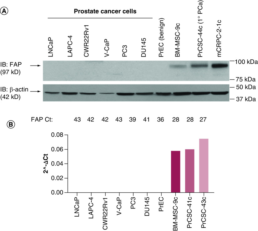
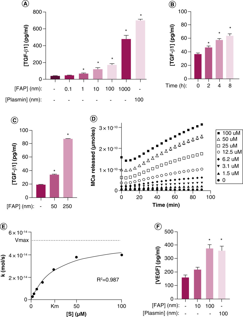
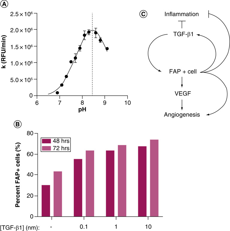
The tumor microenvironment contributes to disease progression through multiple mechanisms, including immune suppression mediated in part by fibroblast activation protein (FAP)-expressing cells. Herein, a review of FAP biology is presented, supplemented with primary data. This includes FAP expression in prostate cancer and activation of latent reservoirs of TGF-β and VEGF to produce a positive feedback loop. This collectively suggests a normal wound repair process subverted during cancer pathophysiology. There has been immense interest in targeting FAP for diagnostic, monitoring and therapeutic purposes. Until recently, this development has outpaced an understanding of the biology; impeding optimal translation into the clinic. A summary of these applications is provided with an emphasis on eliminating tumor-infiltrating FAP-positive cells to overcome stromal barriers to immuno-oncological responses.

Keywords: FAP; TGF-β; fibroblast activation protein; immunotherapy; prostate cancer; stroma; wound healing.


Conflict of interest statement

Financial & competing interests disclosure

The work was supported by Abbvie (C109738FE, [WN Brennen]), Allegheny Health Network-Johns Hopkins University Cancer Research Fund (WN Brennen, DLJ Thorek), Emerson Collective Cancer Research Fund (643396, [WN Brennen, DLJ Thorek]), the Department of Defense (W81XWH-17-1-0528, [WN Brennen]), W81XWH-16-1-0410 (JT Isaacs, SR Denmeade), and the NIH-Prostate SPORE Grant (P50 CA058236, [SR Denmeade, JT Isaacs]). The authors have no other relevant affiliations or financial involvement with any organization or entity with a financial interest in or financial conflict with the subject matter or materials discussed in the manuscript apart from those disclosed.

No writing assistance was utilized in the production of this manuscript.

Figures

Figure 1.
Figure 1.. Fibroblast activation protein expression in epithelial and stromal cells derived from benign and malignant prostate tissue.
(A) FAP immunoblot documenting no FAP expression in benign or malignant prostate epithelial cells. However, FAP is detected on primary BM-MSCs and prostate cancer stromal cells derived from primary (1° PCa) and metastatic castration-resistant prostate cancer lesions. (B) qRT-PCR documenting high levels of FAP mRNA detected in BM-MSCs and prostate cancer stromal cells, but not in prostate cancer epithelial cells. Results confirmed with an independent set of primers. FAP: Fibroblast activation protein; BM-MSC: Bone marrow-derived mesenchymal stem cell.
Figure 2.
Figure 2.. Fibroblast activation protein activates latent reservoirs of TGF-β sequestered in the basement membrane and extracellular matrix.
(A) HUVEC treated with increasing concentrations of exogenous recombinant human FAP (rhFAP) for 2.5 h at 37°C demonstrates a dose-dependent increase in TGF-β1 in the supernatant compared with vehicle-treated controls as measured by ELISA from two independent experiments performed in duplicate. Plasmin used as a positive control. (B) rhFAP (50 nM) releases TGF-β1 from HUVEC-derived basement membrane denuded of cells in a time-dependent manner as determined by ELISA of conditioned media at the indicated time points. Experiment performed in duplicate. (C) FAP releases TGF-β1 from human fibroblast (WPMY-1) derived ECM denuded of cells in a concentration-dependent manner over 24 h at 37°C. Three independent experiments performed in duplicate. (D) FAP-dependent cleavage of a fluorescence-quenched peptide substrate (Mca-EIPESGPSSG-Dnp) designed from the hinge region of LTBP-4 in the presence of FAP (100 nM) at 37°C. No Mca released in the absence of FAP. All experiments performed in triplicate with a representative plot shown here. (E) Representative plot of hydrolysis rate (k) versus substrate concentration ([S]) for the peptide presented in panel D. (F) Increasing concentrations of VEGF detected in conditioned media from human prostate cancer (PC3) cells treated with increasing concentrations of exogenous rhFAP release as measured by ELISA and measured in triplicate. Plasmin used as a positive control. All error bars represent standard error (SE). p-values < 0.05 (*) relative to the untreated controls. ECM: Extracellular matrix; FAP: Fibroblast activation protein.
Figure 3.
Figure 3.. Fibroblast activation protein pH profile and positive feedback loop with TGF-β.
(A) FAP is optimally active under alkaline conditions. The rate of substrate (AP-AFC) hydrolysis (k) was measured in the presence or absence of FAP in a series of Tris buffers at the indicated pH values at 37°C. All samples run in triplicate. (B) TGF-β1 induces FAP expression on human prostate fibroblasts (WPMY-1) in a concentration-dependent manner as determined by flow cytometry. (C) Model of positive feedback loop between FAP+ cells and TGF-β1 regulating angiogenesis and inflammation during wound healing and chronic inflammation with implications for cancer initiation and progression. All error bars represent standard error. FAP: Fibroblast activation protein.

Similar articles

Cited by

References

    1. Brennen WN, Isaacs JT, Denmeade SR. Rationale behind targeting fibroblast activation protein-expressing carcinoma-associated fibroblasts as a novel chemotherapeutic strategy. Mol. Cancer Ther. 11(2), 257–266 (2012). - PMC - PubMed
    1. Caplan AI, Sorrell JM. The MSC curtain that stops the immune system. Immunol. Lett. 2015) (Epub ahead of print). - PubMed
    1. Krueger TE, Thorek DLJ, Meeker AK, Isaacs JT, Brennen WN. Tumor-infiltrating mesenchymal stem cells: drivers of the immunosuppressive tumor microenvironment in prostate cancer? Prostate 79(3), 320–330 (2019). - PMC - PubMed
    1. Costa A, Kieffer Y, Scholer-Dahirel A et al. Fibroblast heterogeneity and immunosuppressive environment in human breast cancer. Cancer Cell 33(3), 463–479 e410 (2018). - PubMed
    1. Lambrechts D, Wauters E, Boeckx B et al. Phenotype molding of stromal cells in the lung tumor microenvironment. Nat. Med. 24(8), 1277–1289 (2018). - PubMed

Publication types

MeSH terms

Substances

LinkOut - more resources