

A tumour suppressive relationship between mineralocorticoid and retinoic acid receptors activates a transcriptional program consistent with a reverse Warburg effect in breast cancer
- PMID: 33148314
- PMCID: PMC7641839
- DOI: 10.1186/s13058-020-01355-x
A tumour suppressive relationship between mineralocorticoid and retinoic acid receptors activates a transcriptional program consistent with a reverse Warburg effect in breast cancer
Abstract
Background: The role of nuclear receptors in both the aetiology and treatment of breast cancer is exemplified by the use of the oestrogen receptor (ER) as a prognostic marker and treatment target. Treatments targeting the oestrogen signalling pathway are initially highly effective for most patients. However, for the breast cancers that fail to respond, or become resistant, to current endocrine treatments, the long-term outlook is poor. ER is a member of the nuclear receptor superfamily, comprising 48 members in the human, many of which are expressed in the breast and could be used as alternative targets in cases where current treatments are ineffective.
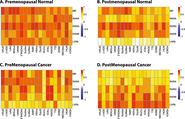
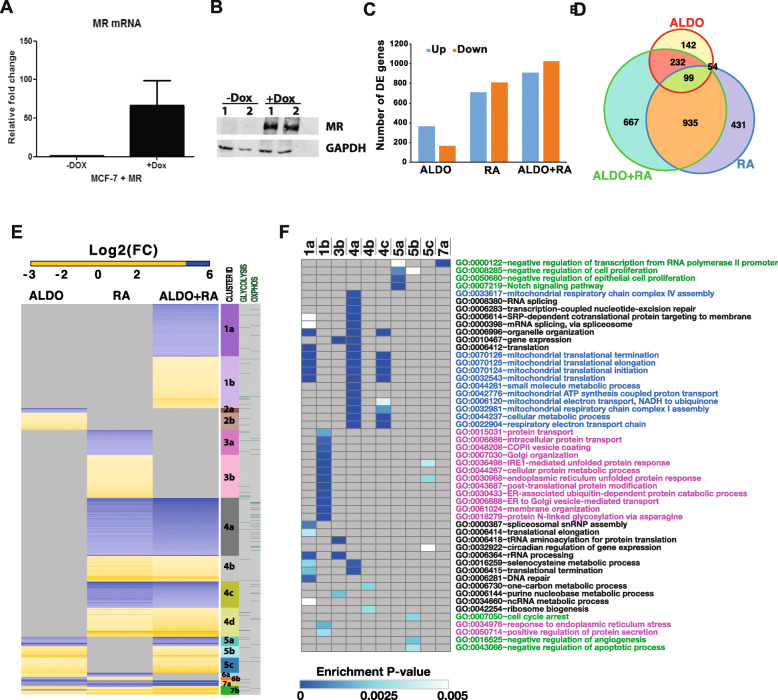
Methods: We used sparse canonical correlation analysis to interrogate potential novel nuclear receptor expression relationships in normal breast and breast cancer. These were further explored using whole transcriptome profiling in breast cancer cells after combinations of ligand treatments.
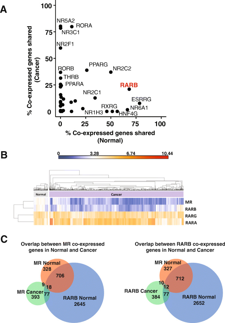
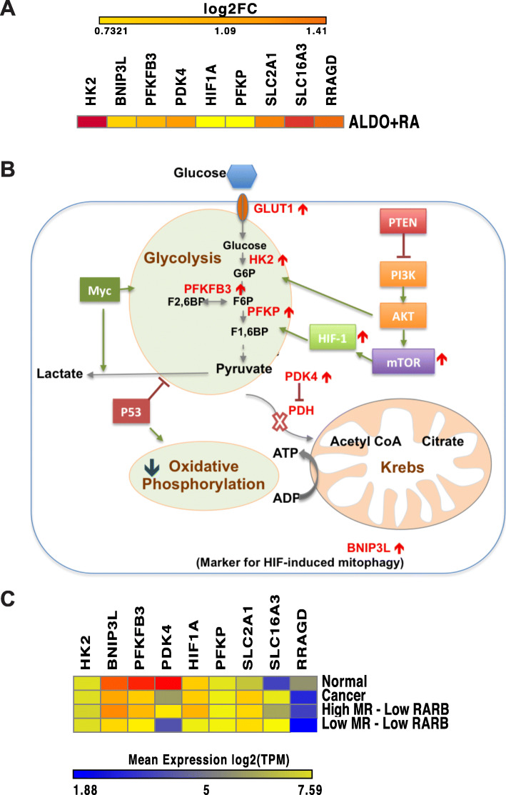
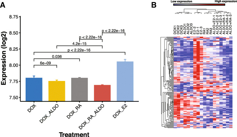
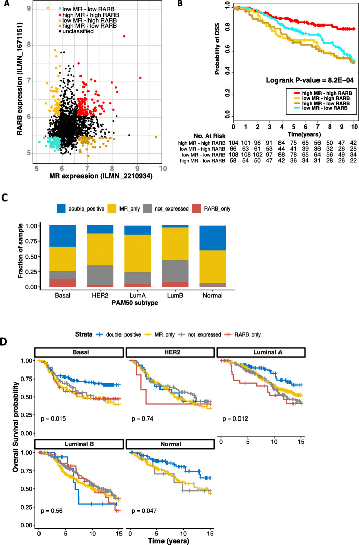
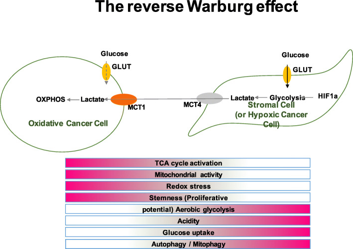
Results: Using this approach, we discovered a tumour suppressive relationship between the mineralocorticoid receptor (MR) and retinoic acid receptors (RAR), in particular RARβ. Expression profiling of MR expressing breast cancer cells revealed that mineralocorticoid and retinoid co-treatment activated an expression program consistent with a reverse Warburg effect and growth inhibition, which was not observed with either ligand alone. Moreover, high expression of both MR and RARB was associated with improved breast cancer-specific survival.
Conclusion: Our study reveals a previously unknown relationship between MR and RAR in the breast, which is dependent on menopausal state and altered in malignancy. This finding identifies potential new targets for the treatment of breast cancers that are refractory to existing therapeutic options.
Keywords: Breast cancer; Gene expression profiling; Mineralocorticoid receptor; Retinoic acid receptor; Warburg effect.
Conflict of interest statement
The authors declare that they have no competing interests.
Figures
Similar articles
-
Retinoic acid receptor alpha expression correlates with retinoid-induced growth inhibition of human breast cancer cells regardless of estrogen receptor status.Cancer Res. 1997 Jul 1;57(13):2642-50. Cancer Res. 1997. PMID: 9205071
-
The N-terminal of the estrogen receptor (ERalpha) mediates transcriptional cross-talk with the retinoic acid receptor in human breast cancer cells.J Steroid Biochem Mol Biol. 2003 Jul;86(1):1-14. doi: 10.1016/s0960-0760(03)00255-3. J Steroid Biochem Mol Biol. 2003. PMID: 12943740
-
Comparison of N-(4-hydroxyphenyl)retinamide and all-trans-retinoic acid in the regulation of retinoid receptor-mediated gene expression in human breast cancer cell lines.Cancer Res. 1996 Mar 1;56(5):1056-62. Cancer Res. 1996. PMID: 8640761
-
Retinoid, retinoic acid receptor beta and breast cancer.Breast Cancer Res Treat. 2002 Nov;76(2):167-73. doi: 10.1023/a:1020576606004. Breast Cancer Res Treat. 2002. PMID: 12452454 Review.
-
Retinoid receptors in human lung cancer and breast cancer.Mutat Res. 1996 Feb 19;350(1):267-77. doi: 10.1016/0027-5107(95)00102-6. Mutat Res. 1996. PMID: 8657191 Review.
Cited by
-
BC-N102 suppress breast cancer tumorigenesis by interfering with cell cycle regulatory proteins and hormonal signaling, and induction of time-course arrest of cell cycle at G1/G0 phase.Int J Biol Sci. 2021 Jul 25;17(12):3224-3238. doi: 10.7150/ijbs.62808. eCollection 2021. Int J Biol Sci. 2021. PMID: 34421361 Free PMC article.
-
Epigenetic regulation of RARB overcomes the radio-resistance of colorectal carcinoma cells via cancer stem cells.J Radiat Res. 2023 Jan 20;64(1):11-23. doi: 10.1093/jrr/rrac060. J Radiat Res. 2023. PMID: 36214504 Free PMC article.
-
The Sin3A/MAD1 Complex, through Its PAH2 Domain, Acts as a Second Repressor of Retinoic Acid Receptor Beta Expression in Breast Cancer Cells.Cells. 2022 Mar 31;11(7):1179. doi: 10.3390/cells11071179. Cells. 2022. PMID: 35406744 Free PMC article.
-
Lactic acid metabolism: gynecological cancer's Achilles' heel.Discov Oncol. 2025 May 2;16(1):657. doi: 10.1007/s12672-025-02364-y. Discov Oncol. 2025. PMID: 40314877 Free PMC article. Review.
-
Stress regulatory hormones and cancer: the contribution of epinephrine and cancer therapeutic value of beta blockers.Endocrine. 2025 May;88(2):359-386. doi: 10.1007/s12020-025-04161-7. Epub 2025 Jan 27. Endocrine. 2025. PMID: 39869294 Review.
References
-
- Jensen EV, Jordan VC. The estrogen receptor: a model for molecular medicine. Clin Cancer Res. 2003;9(6):1980–1989. - PubMed
-
- Gronemeyer H, Gustafsson JA, Laudet V. Principles for modulation of the nuclear receptor superfamily. Nat Rev Drug Discov. 2004;3(11):950–964. - PubMed
-
- Fuller PJ. Novel interactions of the mineralocorticoid receptor. Mol Cell Endocrinol. 2015;408:33–37. - PubMed
-
- Pascual-Le Tallec L, Lombes M. The mineralocorticoid receptor: a journey exploring its diversity and specificity of action. Mol Endocrinol. 2005;19(9):2211–2221. - PubMed
Publication types
MeSH terms
Substances
LinkOut - more resources
Full Text Sources
Medical
