. 2020 Nov 4;21(1):271.
doi: 10.1186/s13059-020-02175-0.

Multiomics profiling of primary lung cancers and distant metastases reveals immunosuppression as a common characteristic of tumor cells with metastatic plasticity

Affiliations

Multiomics profiling of primary lung cancers and distant metastases reveals immunosuppression as a common characteristic of tumor cells with metastatic plasticity

Won-Chul Lee et al. Genome Biol. .

Abstract

Background: Metastasis is the primary cause of cancer mortality accounting for 90% of cancer deaths. Our understanding of the molecular mechanisms driving metastasis is rudimentary.

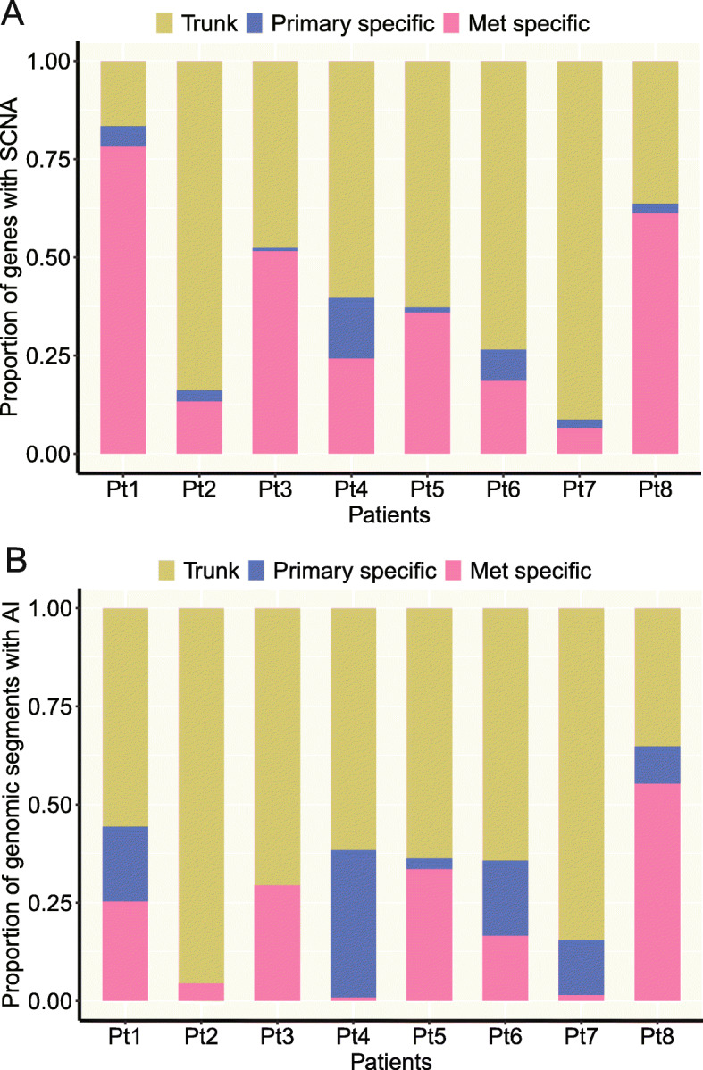
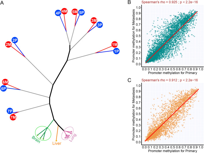
Results: We perform whole exome sequencing (WES), RNA sequencing, methylation microarray, and immunohistochemistry (IHC) on 8 pairs of non-small cell lung cancer (NSCLC) primary tumors and matched distant metastases. Furthermore, we analyze published WES data from 35 primary NSCLC and metastasis pairs, and transcriptomic data from 4 autopsy cases with metastatic NSCLC and one metastatic lung cancer mouse model. The majority of somatic mutations are shared between primary tumors and paired distant metastases although mutational signatures suggest different mutagenesis processes in play before and after metastatic spread. Subclonal analysis reveals evidence of monoclonal seeding in 41 of 42 patients. Pathway analysis of transcriptomic data reveals that downregulated pathways in metastases are mainly immune-related. Further deconvolution analysis reveals significantly lower infiltration of various immune cell types in metastases with the exception of CD4+ T cells and M2 macrophages. These results are in line with lower densities of immune cells and higher CD4/CD8 ratios in metastases shown by IHC. Analysis of transcriptomic data from autopsy cases and animal models confirms that immunosuppression is also present in extracranial metastases. Significantly higher somatic copy number aberration and allelic imbalance burdens are identified in metastases.

Conclusions: Metastasis is a molecularly late event, and immunosuppression driven by different molecular events, including somatic copy number aberration, may be a common characteristic of tumors with metastatic plasticity.

Keywords: DNA methylation; Gene expression; Genomics; Immune profiling; Lung cancer; Metastasis; Multiomics.


Conflict of interest statement

JZ reports research funding from Merck and Johnson & Johnson and personal fees from AstraZeneca, Bristol-Myers Squibb, Johnson & Johnson GenePlus-Beijing Institute, OrigiMed, and Innovent outside the submitted work. KS reports research funding from Boehringer Ingelheim and Rain Therapeutics, and personal fees from AstraZeneca and Boehringer Ingelheim (all outside the submitted work). DLG has served on the scientific advisory committees for AstraZeneca, GlaxoSmithKline, Sanofi, and Janssen and has received research support from Janssen, Takeda, Ribon Therapeutics, Astellas, and AstraZeneca.

Figures

Fig. 1
Fig. 1
Genetic divergence of primary lung tumors and paired distant metastases. For each patient, the left scatter plot shows cancer cell fraction (CCF) values for somatic single nucleotide variants. Variants with different color show the different clones. In the phylogenetic trees of primary (P) and metastatic (M) tumors, trunk and branch lengths are proportional to the number of somatic mutations. Cancer gene mutations are displayed with the trees (oncogene in red and tumor suppressor gene in green)
Fig. 2
Fig. 2
The level of concordance for somatic copy number aberrations (SCNAs) and allelic imbalance (AI). a The proportions of trunk, primary-specific, and metastasis-specific SCNA events. SCNA events were defined at gene level. Specifically, segment log2 ratio means were assigned to genes within each segment with SCNA so each sample would have log2 ratio values of the same number of genes for fair comparison between samples. b The proportions of trunk, primary-specific, and metastasis-specific AI events
Fig. 3
Fig. 3
DNA methylation similarity between primary tumors and paired metastases. a Unsupervised clustering of all samples including normal tissues based on DNA methylation level of all CpG islands (n = 27,000). b Correlation of promoter DNA methylation between primary tumors and metastases for the 1084 genes showing a negative correlation between DNA methylation and gene expression (Spearman’s rank correlation ≤ − 0.5 based on the data in our main cohort). c Correlation of promoter DNA methylation level between primary tumors and metastases for the 521 genes previously reported to be regulated by DNA methylation in NSCLC from the study examining 73 cell lines
Fig. 4
Fig. 4
Differential signaling pathways in metastasis and immunohistochemical assessment of leukocyte antigens. a Unsupervised clustering of gene expression profiles using highly variable genes (standard deviation > 2.0; n = 4139). The complete linkage and 1-correlation distance metric were used. Each row represents a gene, and each column represents a sample. Tumor versus normal: T, tumor; N, normal. Tissue type: P, primary tumor; M, metastasis; PN, adjacent normal lung; MN, metastasis adjacent normal tissue. Organ: L, lung; B, brain; H, liver. b Upregulated and downregulated pathways in metastasis (nominal p < 0.01 and q < 0.25). Pathways in red are upregulated pathways in metastasis, and pathways in blue are downregulated pathways in metastasis. c Comparison of immune cell infiltration between primary NSCLC tumors and paired metastases by immunohistochemistry (IHC) of immune markers (CD3, CD4, CD8, CD20, and PD1). The density was defined as the number of cells positive for each marker per square millimeter. The y axis shows the ratio (log2) of density of each cell type in metastases versus that in paired primary lung cancers
Fig. 5
Fig. 5
Immune cell infiltration in primary lung cancers versus metastases by deconvolution of transcriptomic profiles. a The overall immune cell infiltration was inferred by RNA-seq data using ESTIMATE. bg The immune cell subsets were inferred by deconvolution of RNA-seq data using TIMER. The y axis represents the proportion of each immune cell type in the specimen. h The CD4/CD8 ratio inferred using TIMER. The difference was assessed by the paired-sample Wilcoxon test

References

    1. Chaffer CL, Weinberg RA. A perspective on cancer cell metastasis. Science. 2011;331:1559–1564. doi: 10.1126/science.1203543. - DOI - PubMed
    1. Sporn MB. The war on cancer. Lancet. 1996;347:1377–1381. doi: 10.1016/S0140-6736(96)91015-6. - DOI - PubMed
    1. Nowell PC. The clonal evolution of tumor cell populations. Science. 1976;194:23–28. doi: 10.1126/science.959840. - DOI - PubMed
    1. Valastyan S, Weinberg RA. Tumor metastasis: molecular insights and evolving paradigms. Cell. 2011;147:275–292. doi: 10.1016/j.cell.2011.09.024. - DOI - PMC - PubMed
    1. Yates LR, Campbell PJ. Evolution of the cancer genome. Nat Rev Genet. 2012;13:795–806. doi: 10.1038/nrg3317. - DOI - PMC - PubMed

Publication types