. 2020 Nov 4;12(568):eaay2104.
doi: 10.1126/scitranslmed.aay2104.

Multimodal imaging of bacterial-host interface in mice and piglets with Staphylococcus aureus endocarditis

Affiliations

Multimodal imaging of bacterial-host interface in mice and piglets with Staphylococcus aureus endocarditis

Peter Panizzi et al. Sci Transl Med. .

Abstract

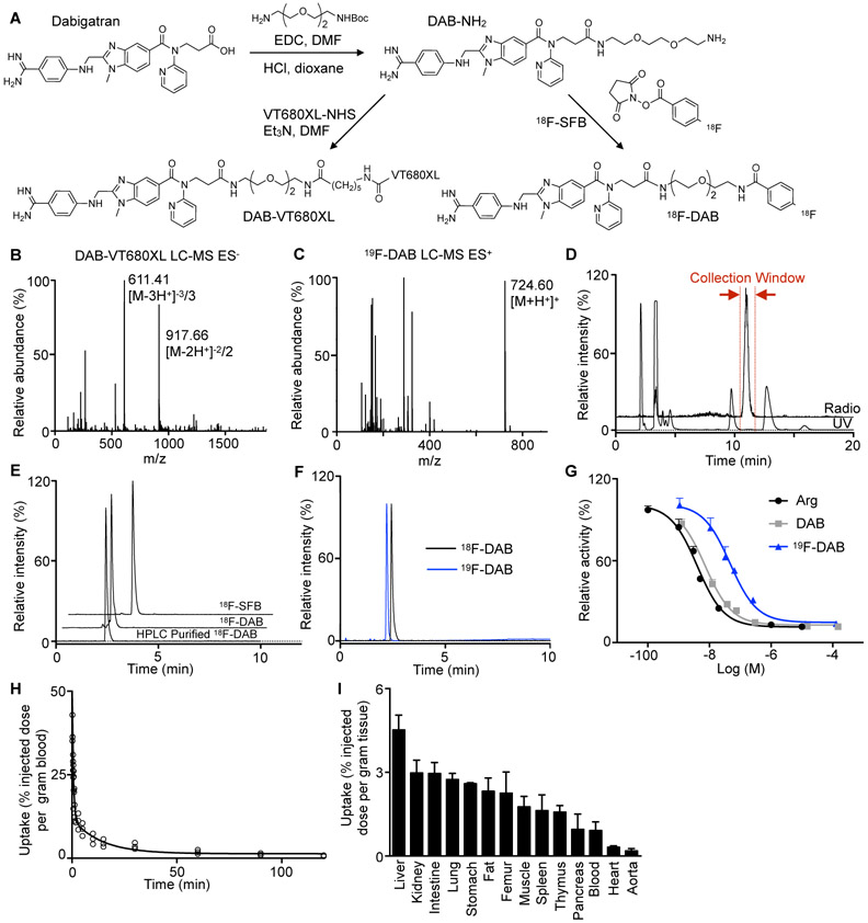
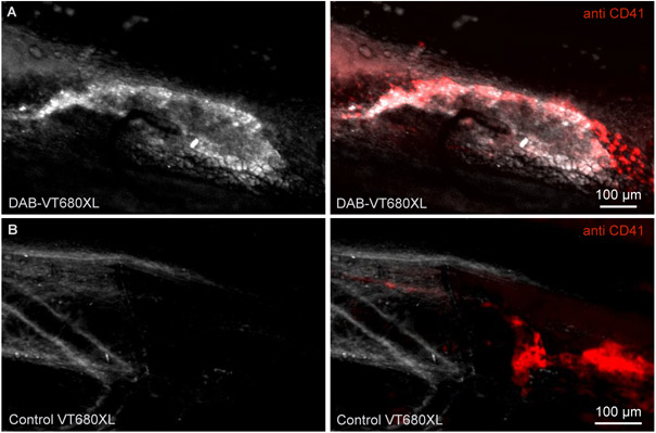
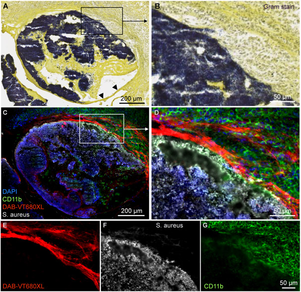
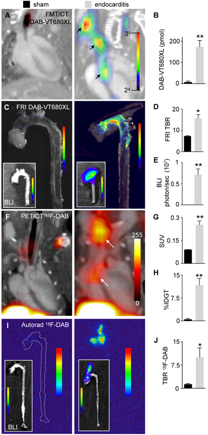
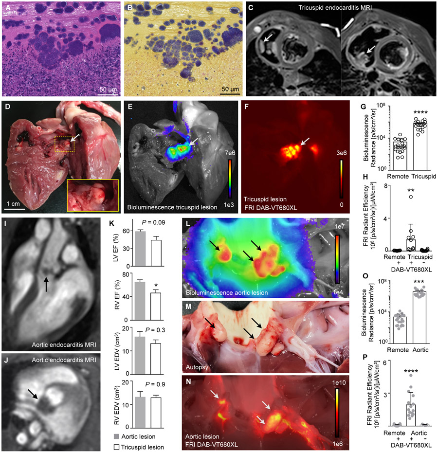
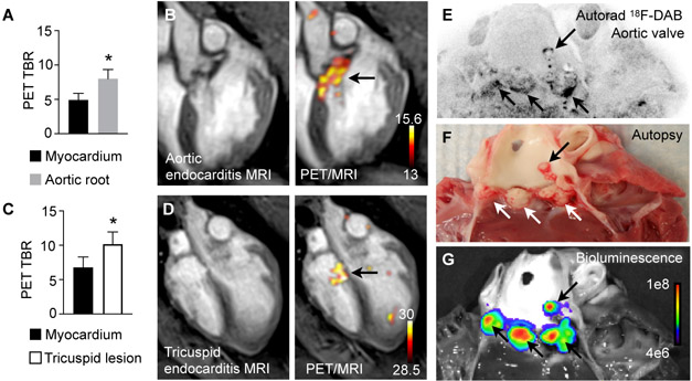
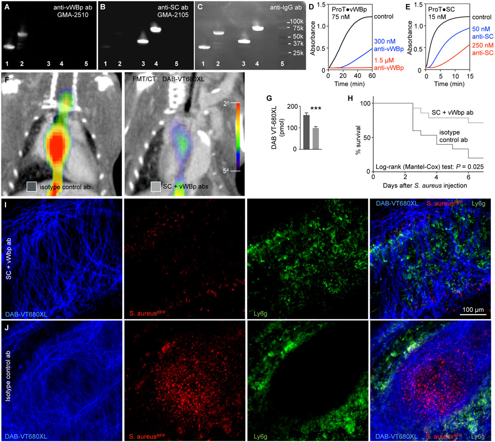
Acute bacterial endocarditis is a rapid, difficult to manage, and frequently lethal disease. Potent antibiotics often cannot efficiently kill Staphylococcus aureus that colonizes the heart's valves. S. aureus relies on virulence factors to evade therapeutics and the host's immune response, usurping the host's clotting system by activating circulating prothrombin with staphylocoagulase and von Willebrand factor-binding protein. An insoluble fibrin barrier then forms around the bacterial colony, shielding the pathogen from immune cell clearance. Targeting virulence factors may provide previously unidentified avenues to better diagnose and treat endocarditis. To tap into this unused therapeutic opportunity, we codeveloped therapeutics and multimodal molecular imaging to probe the host-pathogen interface. We introduced and validated a family of small-molecule optical and positron emission tomography (PET) reporters targeting active thrombin in the fibrin-rich environment of bacterial colonies. The imaging agents, based on the clinical thrombin inhibitor dabigatran, are bound to heart valve vegetations in mice. Using optical imaging, we monitored therapy with antibodies neutralizing staphylocoagulase and von Willebrand factor-binding protein in mice with S. aureus endocarditis. This treatment deactivated bacterial defenses against innate immune cells, decreased in vivo imaging signal, and improved survival. Aortic or tricuspid S. aureus endocarditis in piglets was also successfully imaged with clinical PET/magnetic resonance imaging. Our data map a route toward adjuvant immunotherapy for endocarditis and provide efficient tools to monitor this drug class for infectious diseases.


Figures

Fig. 1.
Fig. 1.. Imaging agent synthesis and validation.
(A) Synthesis scheme of a near infrared fluorescent dabigatran derivative (DAB-VT680XL) and a positron emission tomography fluorine-18 tracer 18F-dabigatran (18F-DAB), both prepared in 2 steps from dabigatran. (B) LC-MS analysis of DAB-VT680XL showing the [M - 2H+]2−/2 (917.66 m/z) and [M - 3H+]3−/3 (611.41 m/z) ions and (C) 19F-DAB showing the [M + H+]+ (724.60 m/z). (D) Preparative HPLC chromatograms (radio, top trace) and ultraviolet (UV) absorbance at 254 nm (bottom trace). The collection window is highlighted in red. (E) Analytical HPLC chromatograms of the 18F-succinimidyl fluorobenzoate (18F-SFB) prosthetic group, 18F-DAB crude conjugation reaction mixture and HPLC purified 18F-DAB. (F) Analytical HPLC chromatograms of 18F-DAB and stable-isotope standard 19F-DAB (blue). Since the two detectors are connected in series, there is a 0.2 min delay between UV and radio signals. (G) Thrombin activity assay with argatroban (Arg), dabigatran (DAB) and 19F-dabigatran (19F-DAB). (H) Clearing 18F-DAB from mouse blood (n=3). The half-life in blood is 2.0±0.6 minutes. (I) Bio-distribution of 18F-DAB in mice (n=3-8). Data are shown as mean ± s.e.m..
Fig. 2.
Fig. 2.. In vivo DAB-VT680XL or control probe binding to thrombi in mice.
(A) Intravital microscopy of a FeCl3-induced thrombosis of the femoral artery 90 min after injection of DAB-VT680XL or (B) an unspecific control fluorochrome (VT680XL). Aggregating platelets are stained with anti-CD41 monoclonal antibody (red). The experiments were repeated twice with the same result (n=3 per group).
Fig. 3.
Fig. 3.. DAB-VT680XL targeting of endocarditic vegetations in mice.
(A) Gram stain of aortic root after inducing endocarditis in mice shows S. aureus in dark purple. Arrow heads indicate aortic valve leaflet. (B) High magnification view of boxed area in (A). (C, D) Adjacent sections of the endocarditic vegetation 90 min after DAB-VT680XL injection. DAPI indicates 4′,6-diamidino-2-phenylindole. DAB-VT680XL imaging signal (E) highlights the intersection of bacterial vegetations (F) with the host. Staining for CD11b (G) illustrates the distribution of myeloid cells, which are unable to enter the bacterial colony. The experiment was repeated twice with the same result.
Fig. 4.
Fig. 4.. Noninvasively imaging mouse endocarditis.
(A) In vivo FMT/CT images and (B) fluorescence quantification in the aortic roots of mice with S. aureus endocarditis (right) and sham controls (left) (n=5-8 mice per group; P = 0.0055). (C) Ex vivo fluorescence reflectance imaging (FRI) and bioluminescence imaging (BLI, insets) illustrate macroscopic co-localization of bacterial colonies with DAB-VT680XL imaging signal. (D) Quantification of fluorescence as target to background ratio (TBR) (n=5-8 mice per group, P = 0.0286). (E) Quantification of BLI signal derived from bioluminescent S. aureus strain Xen29 (n=5-8 mice per group, P = 0.0095). (F) PET/CT imaging after injection of 18F-DAB. Arrows indicate signal from S. aureus vegetations on the aortic valves and the suture in the brachiocephalic artery. (G) In vivo quantification of PET signal in the aortic root (SUV, standardized uptake value, n=6-10 mice per group, data from four separate experiments, P = 0.0095). (H) Ex vivo scintillation counting of aortic roots (%IDGT, percent injected dose per gram tissue, n=5-12 mice per group, P = 0.0052). (I) Autoradiography of dissected aortas. Left panel, sham-operated mouse with absent 18F-DAB signal and absent bioluminescent signal indicating lack of bacteria; right panel with 18F-DAB signal with co-localized bioluminescent bacterial colonies in the ascending aorta (inset). (J) Quantification of autoradiography (TBR, target to background ratio, n=5-7 mice per group, three separate experiments, P = 0.037). Unpaired, two-tailed t-test was used and data are shown as mean ± s.e.m.. *P < 0.05, **P < 0.01.
Fig. 5.
Fig. 5.. S. aureus endocarditis in piglets.
(A) H&E staining of right-sided piglet endocarditis vegetation indicating immune response to the S. aureus Xen 36 infection. (B) Gram staining of an adjacent section showing the pathogen in the piglet heart. (C) Short axis stacks of black blood MRI from a pig 10 days after induction of right-sided endocarditis. Arrows indicate vegetations. (D) View of the open right ventricle in piglet with right-sided endocarditis. Inset shows magnified view. (E) Bioluminescence image of (D) illustrates location of Xen 36 S. aureus. (F) Fluorescence reflectance image (FRI) of (D) and (E) indicates DAB-VT680XL imaging signal after intravenous injection of the near infrared imaging agent. (G) Ex vivo quantification of bacterial bioluminescence from multiple vegetations of 12 piglets with right-sided endocarditis. Unpaired, two-tailed t-test was used and data are shown as mean ± s.e.m., ****P < 0.0001. (H) FRI in a DAB-VT680XL-injected subset of piglet demonstrates accumulation of the imaging probe (n=3 piglets per group, one-way ANOVA for multiple comparison, Barlett’s test, **P < 0.01. (I) Long and (J) short axis MRI of piglet after induction of left-sided endocarditis. (K) Cardiac MRI-derived ejection fraction (EF) of the right and left ventricle (RV and LV, respectively) and the end-diastolic volumes (EDV), comparing left- with right-sided endocarditis (n=6 piglets with tricuspid right-sided and n=4 with aortic left-sided disease, two tailed t test, *P < 0.05). (L) Bioluminescence imaging demonstrates bacterial infection, with signal arising from vegetations (arrows) identified on autopsy (M) and FRI (N). (O) Ex vivo quantification of bacterial bioluminescence in 8 piglets with left-sided endocarditis, two-tailed t test., ***P < 0.001. (P) Quantification of fluorescence signal arising from aortic valve vegetations (n=5 piglets, one-way ANOVA for multiple comparison with ****P < 0.0001, Barlett’s test).
Fig. 6.
Fig. 6.. PET/MR imaging S. aureus endocarditis in piglets.
(A) Left-sided endocarditis target to background ratio (TBR) of PET signal, calculated with either myocardium (control) or vegetation-bearing aortic root as target and the skeletal muscle as background (n=3 piglets, two-sided Student’s t test, *P < 0.05). (B) PET/MR images illustrating the imaging signal in aortic valve with endocarditis lesion (arrow). Color scale depicts becquerels/mL. (C) Right-sided endocarditis TBR, calculated as above (n=3 piglets, two-sided Student’s t test, *P < 0.05). (D) PET/MR images illustrating the imaging signal in tricuspid valve endocarditis lesion (arrow). Color scale depicts becquerels/mL. (E) Ex vivo autoradiography of aortic valve indicates radioactive signal in the aortic valve vegetations, which were verified on autopsy (F) and a source of bioluminescence (G) arising from S. aureus (arrows).
Fig. 7.
Fig. 7.. Immunotherapy neutralizing virulence factors disrupts vegetations and improves survival.
(A-C) Specificity of monoclonal antibodies by immunoblotting against vWBp-(1-263) in lane 1; vWBp-(1-474) in lane 2; SC-(1-325) in lane 3; SC-(1-660) in lane 4. Lane 5 contains protein standards with the indicated molecular weights. The indicated antibodies, in panel A GMA-2510 monoclonal [anti-von Willebrand factor-binding protein (anti-vWBp)] and in panel B GMA-2105 monoclonal [anti-staphylocoagulase (anti-SC)], are specific for their respective targets. Panel C shows probing for total mouse IgG (anti-IgG polyclonal against both the heavy and light chains of murine IgG) and reflects the bound antibodies shown in panels A and B. (D) Increase in turbidity as measured by absorbance change at 450 nm for mixtures of 1.5 mg/mL fibrinogen and 75 nM prothrombin complexed to vWBp-(1-263) (ProTvWBp) complex in the absence of GMA-2510 antibody (anti-vWBp Ab; black line), in the presence of 300 nM anti-vWBp Ab (blue line) or 1.5 μM anti-vWBp Ab (red line). (E) Similar reactions for 15 nM prothrombin complexed to SC-(1-325) (ProTSC) are shown in the absence of GMA-2105 antibody (anti-SC Ab; black line), in the presence of 50 nM anti-SC ab (blue line) or 250 nM anti-SC ab (red line). (F) In vivo FMT/CT images of S. aureus endocarditis in mice after injection of DAB-VT680XL treated with either isotype control antibody or antibodies neutralizing SC and vWBp. (G) In vivo fluorescence quantitation of vegetation thrombin in aortic roots following DAB-VT680XL injection (n=10-12 mice per group, three separate experiments; unpaired, two-sided t-test was used and data are shown as mean ± s.e.m. ***P < 0.001). (H) Kaplan-Meier survival curves of S. aureus endocarditis mice treated with isotype control antibody or combination therapy with anti-SC and anti-vWBp antibodies (n=15 mice per group, mice received a single intraperitoneal injection of their respective antibody treatment 6 hrs post surgery). (I) Intravital microscopy of femoral S. aureus vegetation 24 hours after intravenous injection of S. aureusRFP+ and combination treatment with both anti-SC and anti-vWBp or (J) isotype control antibody. In vivo DAB-VT680 microscopy of the vegetation wall surrounding RFP+ bacteria and Ly6g+ neutrophils.

Similar articles

Cited by

References

    1. Tong SY, Davis JS, Eichenberger E, Holland TL, Fowler VG, Staphylococcus aureus infections: epidemiology, pathophysiology, clinical manifestations, and management. Clin Microbiol Rev. 28(3),603–61 (2015). - PMC - PubMed
    1. Cahill TJ, Baddour LM, Habib G, Hoen B, Salaun E, Pettersson GB, Schäfers HJ, Prendergast BD, Challenges in Infective Endocarditis. J Am Coll Cardiol. 69(3),325–344 (2017). - PubMed
    1. Loeb L, The influence of certain bacteria on the coagulation of the blood. J Med Res. 10(3):,407–19 (1903). - PMC - PubMed
    1. Friedrich R, Panizzi P, Fuentes-Prior P, Richter K, Verhamme I, Anderson PJ, Kawabata S, Huber R, Bode W, Bock PE, Staphylocoagulase is a prototype for the mechanism of cofactor-induced zymogen activation. Nature. 425(6957),535–9 (2003). - PubMed
    1. Kroh HK, Panizzi P, Bock PE, Von Willebrand factor-binding protein is a hysteretic conformational activator of prothrombin. Proc Natl Acad Sci U S A. 106(19),7786–91 (2009). - PMC - PubMed

Publication types