. 2020 Nov 4;6(45):eabb5924.
doi: 10.1126/sciadv.abb5924. Print 2020 Nov.

Small-molecule sequestration of amyloid-β as a drug discovery strategy for Alzheimer's disease

Affiliations

Small-molecule sequestration of amyloid-β as a drug discovery strategy for Alzheimer's disease

Gabriella T Heller et al. Sci Adv. .

Abstract

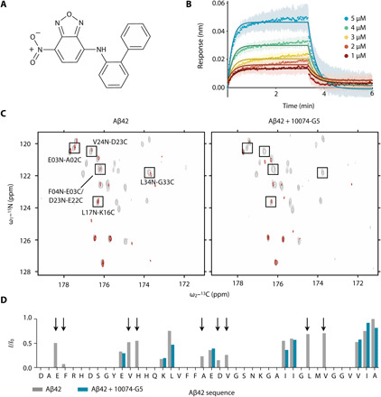
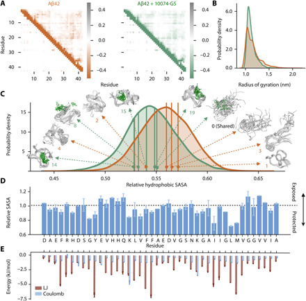
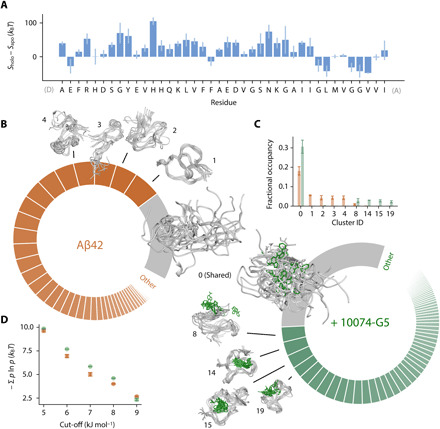
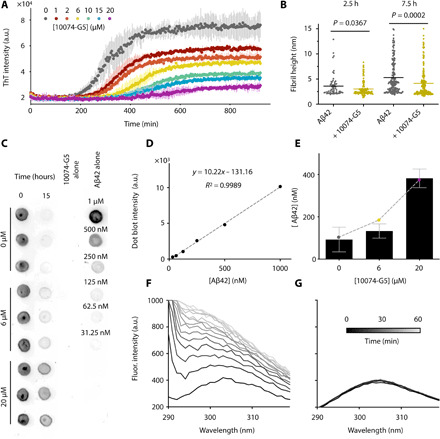
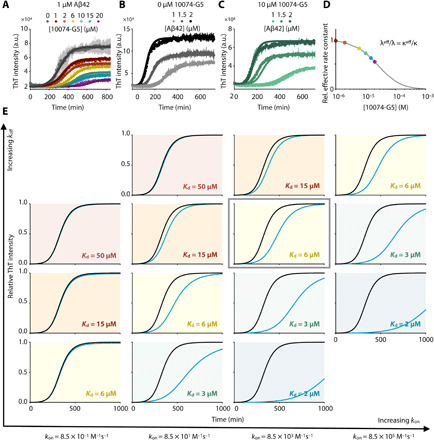
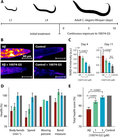
Disordered proteins are challenging therapeutic targets, and no drug is currently in clinical use that modifies the properties of their monomeric states. Here, we identify a small molecule (10074-G5) capable of binding and sequestering the intrinsically disordered amyloid-β (Aβ) peptide in its monomeric, soluble state. Our analysis reveals that this compound interacts with Aβ and inhibits both the primary and secondary nucleation pathways in its aggregation process. We characterize this interaction using biophysical experiments and integrative structural ensemble determination methods. We observe that this molecule increases the conformational entropy of monomeric Aβ while decreasing its hydrophobic surface area. We also show that it rescues a Caenorhabditis elegans model of Aβ-associated toxicity, consistent with the mechanism of action identified from the in silico and in vitro studies. These results illustrate the strategy of stabilizing the monomeric states of disordered proteins with small molecules to alter their behavior for therapeutic purposes.


Figures

Fig. 1
Fig. 1. Characterization of the interaction of 10074-G5 with monomeric Aβ42.
(A) Structure of biphenyl-2-yl-(7-nitro-benzo[1,2,5]oxadiazol-4-yl)-amine, also known as 10074-G5. (B) Biolayer interferometry measurements showing the dose-dependent binding of 10074-G5 to an Aβ42-functionalized surface at various concentrations of the added compound. The curves were corrected for baseline drift. Raw data are shown in fig. S1A. Control curves showing negligible nonspecific binding are shown in fig. S1 (B and D). Global fitting to simple one-phase association and dissociation equations yields association (kon) and dissociation (koff) rates to be 8.5 × 10³ ± 0.2 × 10³ M−1 s−1 and 4.7 × 10⁻² ± 2 × 10⁻⁴ s−1, respectively, corresponding to a binding dissociation constant (Kd) of 6 μM. For this fit, all five curves were constrained to single, shared kon and koff values. The global R2 for the fits is 0.98. (C) 2D HN–BESTCON spectra in the absence (left) and presence (right) of 1:2 Aβ42:10074-G5 with (red) and without (gray) selective water presaturation, performed at 15°C. (D) Quantification of the relative I/I0 intensities from (C) shows that the peptide amide groups are more exposed to solvent in the presence of 10074-G5 (blue) as compared to its absence (gray). Arrows highlight regions along the sequence in which signals are detectable in the absence of the compound, but not in its presence, thus suggesting that 10074-G5 increases the solvent exposure of specific regions of Aβ42. ppm, parts per million.
Fig. 2
Fig. 2. Metadynamic metainference simulations characterize the dynamic binding and show how 10074-G5 promotes Aβ42 conformations with less hydrophobicity.
(A) Metadynamics metainference simulations demonstrate that inter-residue contact maps for Lennard-Jones (LJ) (top right) and Coulomb (top left) potentials for the unbound (orange) and the bound (green) structural ensembles of Aβ42 with 10074-G5 are highly similar. (B) Radii of gyration for the unbound and bound structural ensembles are also highly similar (shown using kernel density estimates of 35,000 points each sampled based on metadynamics weights using a Gaussian kernel). (C) Relative hydrophobic solvent accessible surface area (SASA) of Aβ42 (total hydrophobic area over total surface area) of the bound and unbound ensembles, showing that 10074-G5 decreases the relative exposed hydrophobicity of Aβ42. The holo ensemble was calculated only on the protein surface but accounts for the presence of the compound. Data are shown using kernel density estimates as described in (B). Some of the representative structures from these distributions are shown. Numbers indicate cluster IDs shown in Fig. 3. (D) Ratio (bound/unbound) of the ensemble-averaged, total SASA per residue showing regions of Aβ42 that become more exposed or protected in the presence of 10074-G5. SASAs of the bound ensemble were calculated on the protein in the presence of 10074-G5. (E) Ensemble-averaged, residue-specific LJ and Coulomb interaction energies show that 10074-G5 has strong interactions with aromatic and charged residues. Error bars represent SDs between first and second halves of the analyzed trajectories in (D) and (E).
Fig. 3
Fig. 3. 10074-G5 increases the conformational entropy of Aβ42.
(A) Residue-specific differences in the conformational entropies, S, between the holo and apo ensembles, estimated from normalized, two-dimensional (2D) Ramachandran histograms (100 × 100 bins) of each residue using S = − Σ b ln b where b is the occupancy of a given bin. (B) Donut plots quantifying the conformational states of Aβ42 in the unbound (left, orange) and bound (right, green) simulations. Clustering was performed on concatenated trajectories, considering only Aβ42. Inter-residue contact maps based on the Lennard-Jones potential were used as input for GROMOS clustering (34). The cut-off value is 8.5 kJ mol−1. Each slice represents a distinct state. The simulations share one major state (cluster 0, gray), which comprises 18 and 31% of the unbound and bound ensembles, respectively. (C) Convergence of the five most populated clusters. Bar plot shows fractional cluster occupancies for the unbound (orange) and bound (green) simulations. Fractional cluster occupancies were calculated on 35,000 frames for each concatenated trajectory sampled based on metadynamics weights. (D) The conformational entropy of Aβ42, estimated via Gibbs entropy, is consistently higher in the 10074-G5–bound form of the peptide for several clustering cut-off values. The conformational entropy was calculated such that the weights, P, of each state correspond to the fractional occupancy as determined by the GROMOS clustering algorithm (34). Error bars represent ± SDs of values calculated from the first and second halves of the simulations in (C) and (D).
Fig. 4
Fig. 4. 10074-G5 sequesters monomeric Aβ42 and inhibits its aggregation.
(A) ThT measurements using 1 μM Aβ42 show concentration-dependent effects of 10074-G5 on Aβ42 aggregation. Measurements were taken in triplicate. The concentration of DMSO was held constant across all samples. (B) Distributions of cross-sectional heights of 1 μM Aβ42 fibrils at 2.5 hours (n ≥ 60) and 7.5 hours (n ≥ 200) formed with and without 6 μM 10074-G5, from single-molecule analyses of AFM maps (fig. S7). Lines indicate means. P values were determined by unpaired, two-tailed Student’s t test. Fibrillar aggregates formed in the presence of 10074-G5 have smaller cross-sectional diameters than those formed in its absence. (C) Dot blot of soluble Aβ42 before and after the aggregation of 1 μM Aβ42 with and without 10074-G5 using the W0-2 antibody indicates sequestration of soluble Aβ42. Blotting was performed in triplicate. Fit (D) and quantification (E) used to estimate the concentration of soluble Aβ42 remaining at the end of the aggregation reaction from (C). The dashed curve in (E) represents fit to eq. S14, describing the equilibrium concentration of unreacted monomer from a competitive binding of free monomers to fibril ends and inhibitor. Using this simple fit, we determined the fitted affinity of 10074-G5 for the soluble material to be Kd = 7 ± 1 μM. Intrinsic fluorescence profiles of Tyr10 of 5 μM Aβ42 in the absence (F) and presence (G) of 1:1 10074-G5 over 1 hour show that 10074-G5 delays an increase in fluorescence, suggesting that 10074-G5 inhibits early aggregation events including oligomerization and multimerization. Error bars represent ± SDs in (A) and (E).
Fig. 5
Fig. 5. 10074-G5 inhibits Aβ42 aggregation primarily by monomer sequestration.
(A) Global fit of ThT kinetic curves to a monomer sequestration model (eq. S12), in which 10074-G5 affects the aggregation by binding free monomers. Measurements are those shown in Fig. 4A. The theoretical curves are obtained using eq. S11 with unperturbed kinetic obtained from (B) leaving Kd as the only global fitting parameter (eq. S15). The global fit yields Kd = 40 μM. Global fits to eq. S15 were performed on normalized data to extract changes in the rate parameters in the presence of 10074-G5. (B) Global fit to eq. S11 and eq. S12 of ThT kinetic traces of the aggregation reaction for increasing concentrations of Aβ42 (1, 1.5, and 2 μM) in the absence of 10074-G5. Measurements were taken in duplicate or triplicate. (C) Overlay of theoretical kinetic curves from (A) with independent ThT kinetic traces of the aggregation reaction for increasing concentrations of Aβ42 (1, 1.5, and 2 μM) in the presence of 10 μM 10074-G5. Solid curves are predictions of the kinetic monomer sequestration model using the same rate parameters and inhibitor binding constant as in (A) and no fitting parameters. Measurements were taken in duplicate or triplicate. (D) Effective rates of aggregate proliferation through primary (λ) and secondary (κ) nucleation in the presence of varying concentrations of 10074-G5 determined using the global fit in (A). Error bars represent ± SDs in (A to C). (E) Phase diagram illustrating numerical solutions to the kinetic equations for different kon and koff rates. Curves represent kinetic aggregation traces of 1 μM Aβ42 in the absence (black) and presence of 2 μM compound (blue). Diagonals correspond to constant values of Kd. The values of k+kn and k2k+ are the same as those shown in (A).
Fig. 6
Fig. 6. 10074-G5 is effective in reducing functional impairment in a C. elegans model of Aβ42 toxicity.
(A) Treatment profile used for the C. elegans experiments. (B) NIAD-4 staining of C. elegans aggregates in the presence and absence of 5 μM 10074-G5. (C) Quantification of NIAD-4 intensity shown in (B). n ≥ 9. (D) Health scores (%) for the rate of body bends, speed of movement, moving percent, and magnitude of body bends at day 6 of adulthood. The colors are the same as those shown in (C). Scores are normalized to the N2 control strain (gray). n = 150. (E) Combined total health scores [average of health scores in (D)]. In all panels, error bars represent ± SEM. P values were determined by two-tailed Student’s t test. 10074-G5 shows minimal movement effects on wild-type C. elegans (fig. S10).

Similar articles

Cited by

References

    1. Cummings J., Lee G., Ritter A., Zhong K., Alzheimer’s disease drug development pipeline: 2019. Alzheimers Dement. 5, 272–293 (2019). - PMC - PubMed
    1. Knowles T. P., Vendruscolo M., Dobson C. M., The amyloid state and its association with protein misfolding diseases. Nat. Rev. Mol. Cell Biol. 15, 384–396 (2014). - PubMed
    1. Cohen S. I. A., Linse S., Luheshi L. M., Hellstrand E., White D. A., Rajah L., Otzen D. E., Vendruscolo M., Dobson C. M., Knowles T. P. J., Proliferation of amyloid-β42 aggregates occurs through a secondary nucleation mechanism. Proc. Natl. Acad. Sci. U.S.A. 110, 9758–9763 (2013). - PMC - PubMed
    1. Hardy J., Selkoe D. J., The amyloid hypothesis of Alzheimer’s disease: Progress and problems on the road to therapeutics. Science 297, 353–356 (2002). - PubMed
    1. Haass C., Selkoe D. J., Soluble protein oligomers in neurodegeneration: Lessons from the Alzheimer’s amyloid β-peptide. Nat. Rev. Mol. Cell Biol. 8, 101–112 (2007). - PubMed

Publication types

MeSH terms