. 2021 Jan 13;95(3):e01956-20.
doi: 10.1128/JVI.01956-20. Print 2021 Jan 13.

Chikungunya Virus Replication Rate Determines the Capacity of Crossing Tissue Barriers in Mosquitoes

Affiliations

Chikungunya Virus Replication Rate Determines the Capacity of Crossing Tissue Barriers in Mosquitoes

Fernando Merwaiss et al. J Virol. .

Abstract

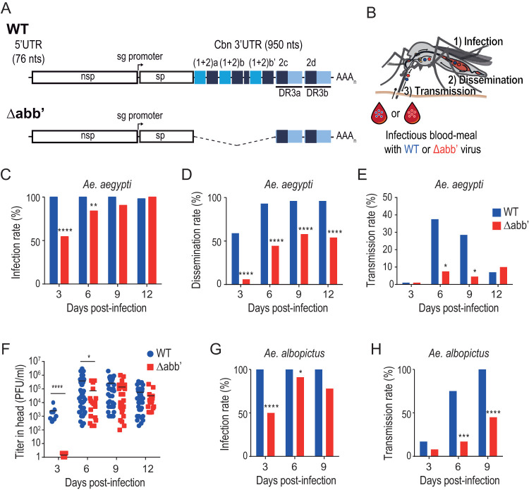
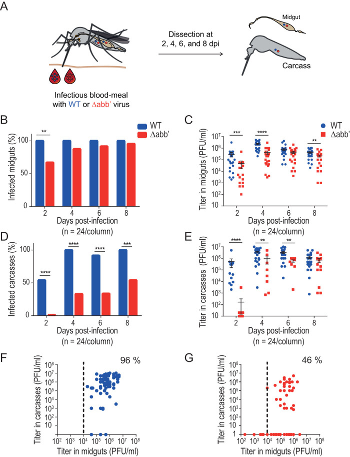
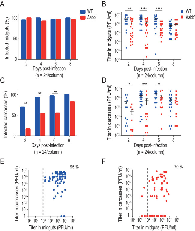
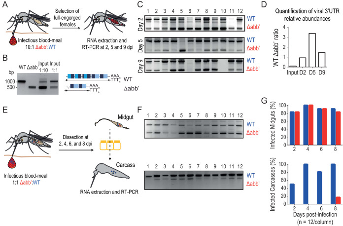
Chikungunya virus (CHIKV) is a reemerging and rapidly spreading pathogen transmitted by mosquitoes. The emergence of new epidemic variants of the virus is associated with genetic evolutionary traits, including duplication of repeated RNA elements in the 3' untranslated region (UTR) that seemingly favor transmission by mosquitoes. The transmission potential of a given variant results from a complex interplay between virus populations and anatomical tissue barriers in the mosquito. Here, we used the wild-type CHIKV Caribbean strain and an engineered mutant harboring a deletion in the 3' UTR to dissect the interactions of virus variants with the anatomical barriers that impede transmission during the replication cycle of the virus in Aedes mosquitoes. Compared to the 3'-UTR mutant, we observed that the wild-type virus had a short extrinsic incubation period (EIP) after an infectious blood meal and was expectorated into mosquito saliva much more efficiently. We found that high viral titers in the midgut are not sufficient to escape the midgut escape barrier. Rather, viral replication kinetics play a crucial role in determining midgut escape and the transmission ability of CHIKV. Finally, competition tests in mosquitoes coinfected with wild-type and mutant viruses revealed that both viruses successfully colonized the midgut, but wild-type viruses effectively displaced mutant viruses during systemic infection due to their greater efficiency of escaping from the midgut into secondary tissues. Overall, our results uncover a link between CHIKV replication kinetics and the effect of bottlenecks on population diversity, as slowly replicating variants are less able to overcome the midgut escape barrier.IMPORTANCE It is well established that selective pressures in mosquito vectors impose population bottlenecks for arboviruses. Here, we used a CHIKV Caribbean lineage mutant carrying a deletion in the 3' UTR to study host-virus interactions in vivo in the epidemic mosquito vector Aedes aegypti We found that the mutant virus had a delayed replication rate in mosquitoes, which lengthened the extrinsic incubation period (EIP) and reduced fitness relative to the wild-type virus. As a result, the mutant virus displayed a reduced capacity to cross anatomical barriers during the infection cycle in mosquitoes, thus reducing the virus transmission rate. Our findings show how selective pressures act on CHIKV noncoding regions to select variants with shorter EIPs that are preferentially transmitted by the mosquito vector.

Keywords: 3′ UTR; alphavirus; arthropod vectors; bottlenecks; extrinsic incubation period.


Figures

FIG 1
FIG 1
Extrinsic incubation period of wild-type and Δabb′ mutant CHIKVs in Aedes mosquitoes. (A) Schematic representation of the genomes of wild-type (WT) and Δabb′ mutant viruses. The Δabb′ mutant bears a deletion of the first 500 nucleotides of the 3′ UTR. (B) Extrinsic incubation period of WT and Δabb′ CHIKVs. Mosquitoes were blood fed with 106 PFU/ml of WT or Δabb′ mutant viruses, and the presence of virus was analyzed in the body (as a proxy of the infection rate), the head (as a proxy of the rate of dissemination to salivary glands), and the saliva (indicative of the transmission rate) at different times postinfection. (C to E) Bar graphs showing infection, dissemination, and transmission rates of WT and Δabb′ viruses in infected Aedes aegypti mosquitoes. (C) The infection rate was calculated as the percentage of infected mosquito bodies at each time point. (D) The dissemination rate was scored as the number of infected mosquito heads over the number of infected bodies. (E) The transmission rate was measured as the ratio between the number of mosquito saliva samples with detectable virus and the number of mosquitoes in which dissemination was successful. Bars for infection, dissemination, and transmission rates represent cumulative data from two independent experiments (n = 48). Data were analyzed by Fisher’s exact test. (F) Dot plot showing mean viral titers and standard deviations (SD) of WT and Δabb′ viruses in the heads of infected mosquitoes. Infectious virus titers were measured in the heads of mosquitoes displaying positive CPE at each time point by plaque assays in Vero cells. Data represent the titers in individual mosquitoes. Statistics were performed by a Mann-Whitney U test. (G and H) Infection and dissemination rates in Aedes albopictus mosquitoes. Bar graphs for infection (G) and dissemination (H) rates are shown (n = 24). Data were analyzed by Fisher’s exact test.
FIG 2
FIG 2
Δabb′ mutant CHIKV is impaired in escaping the midgut. (A) Midgut escape barrier assay. Mosquitoes were blood fed with 106 PFU/ml of wild-type (WT) or Δabb′ mutant CHIKV and dissected from days 2 to 8 to separate midguts and carcasses. Infection rates and viral titers were measured in each sample. dpi, days postinfection. (B) Bar graph showing midgut infection rates. Data represent the percentages of infected mosquito midguts at each time point. (C) Dot plot showing mean viral titers and SD of WT and Δabb′ viruses in midguts of infected mosquitoes. Virus titers in midgut extracts scored positive by a CPE assay were measured by a plaque assay. Data represent titers of individual midguts. (D) Bar graph showing carcass infection rates. Data represent the percentages of infected carcasses at different times after blood feeding and reflect virus dissemination efficiencies. (E) Dot plot showing mean viral titers and SD of WT and Δabb′ viruses in carcasses of infected mosquitoes. Virus titers in carcass extracts were measured by plaque assays. Data represent titers in individual carcasses. (F and G) Scatterplots of viral titers in the midgut versus carcass for individual mosquitoes from the fourth to eighth days postinfection. The dotted line indicates the threshold titer needed to leave the midgut, which was set at 104 PFU/ml. The percentage of mosquitoes above this threshold with disseminated infection was measured for wild-type (F) and mutant (G) viruses. Statistics on infection rates were performed by Fisher’s exact test on cumulative data (n = 24) from two independent experiments. Statistics on viral titers were performed by a Mann-Whitney U test.
FIG 3
FIG 3
Increasing the infectious dose decreases the midgut escape barrier effect. Mosquitoes were blood fed with 5 × 106 PFU/ml wild-type (WT) or Δabb′ mutant CHIKV and dissected from days 2 to 8 to separate midguts and carcasses. Infection rates and viral titers were measured in each sample. (A) Bar graph showing midgut infection rates. (B) Dot plot showing mean viral titers and SD of WT and Δabb′ viruses in midguts of infected mosquitoes. (C) Bar graph showing carcass infection rates. (D) Dot plot showing mean viral titers and SD of WT and Δabb′ viruses in carcasses of infected mosquitoes. (E and F) Scatterplot of viral titers in the midgut versus carcass for wild type (E) and mutant (F) viruses. Statistics on infection rates were performed by Fisher’s exact test on cumulative data (n = 24) from two independent experiments. Statistics on viral titers were performed by a Mann-Whitney U test.
FIG 4
FIG 4
Salivary glands impose a tight barrier to CHIKV transmission. (A) Intrathoracic injections of A. aegypti mosquitoes with wild-type (WT) and Δabb′ CHIKVs. In order to bypass the midgut barrier, A. aegypti mosquitoes were intrathoracically injected with 2,500 PFU of WT or mutant virus. (B) Bar graph showing infection rates in bodies after intrathoracic injection of viruses. The infection rate was calculated as the percentage of mosquitoes with virus presence in the body at different times postinjection. (C) Dot plot showing mean viral titers and SD in the bodies of intrathoracically injected mosquitoes. For the viral titers, statistics were performed by a Mann-Whitney U test. (D) Bar graph showing transmission rates after intrathoracic injection of viruses. The transmission rate was calculated as the percentage of mosquitoes with virus presence in the saliva at different times postinjection.
FIG 5
FIG 5
Wild-type CHIKV has a fitness advantage over Δabb′ CHIKV to cross the midgut escape barrier. (A) Experimental setup of wild-type (WT) versus Δabb′ competitions in Aedes aegypti mosquitoes. Mosquitoes were offered an infectious blood meal containing a mixture of WT and Δabb′ viruses in a 1:10 ratio (106 PFU/ml). Total RNA was purified from individual mosquitoes at different time points postinfection, and the presence of WT and Δabb′ 3′ UTRs was assessed. (B) The RT-PCR product of the RNA extracted from the infectious blood meal containing wild-type and Δabb′ viruses in 1:1 and 1:10 ratios was resolved alongside fragments corresponding to wild-type and Δabb′ 3′ UTRs for reference. (C) Agarose gel electrophoresis of 3′-UTR amplification products from individual mosquitoes. The presence of WT and Δabb′ viruses was assessed by RT-PCR and agarose gel electrophoresis on 12 individual mosquitoes at three different times after the blood meal. (D) Bar graph showing the ratio of WT to Δabb′ 3′ UTRs in the input and in mosquito individuals during the time course of the experiment. Bars represent the average ratios of intensities for the bands corresponding to the products of amplification of WT and Δabb′ 3′ UTRs in individual mosquitoes at each time point. (E) Competition assays to assess the ability of WT and Δabb′ CHIKVs to cross the midgut escape barrier. Infectious blood feeding of A. aegypti mosquitoes was performed with blood containing a mixture of both viruses at a 1:1 ratio (106 PFU/ml). At different times postinfection, the midgut and carcass were dissected, total RNA was extracted, and the presence of virus was evaluated by RT-PCR as described above. (F) Representative agarose gels showing the products of amplification from midgut (top) and carcass (bottom) samples of 12 individual mosquitoes at 4 days postinfection. (G, top) Bar graph showing the presence of WT and Δabb′ viruses in the midgut as a function of time. Bars represent the percentages of midguts where WT and Δabb′ viruses were detected. (Bottom) Bar graph showing the presence of WT and/or Δabb′ viruses in carcasses as a function of time. Bars represent the percentages of carcasses where WT and/or Δabb′ viruses were detected.
FIG 6
FIG 6
Model for the effect of the viral growth rate on the ability to cross barriers during the infectious cycle in mosquitoes. The infection rate in Aedes mosquitoes (midgut infection barrier) is almost 100%, regardless of the virus growth rate. Within midgut cells, wild-type (WT) CHIKV replicates and reaches the necessary threshold (>10,000 PFU) to cross the midgut escape barrier and spread into secondary tissues. A slow-growing virus accomplishes leaving the midgut at later times, and it spreads to secondary tissues in only 50% of individuals. WT disseminated viruses colonize the salivary glands and are successfully secreted into the saliva in 40% of individuals. Secretion into the saliva of mutant viruses is achieved in only 10% of mosquitoes with disseminated infection. The outcome is a longer EIP and a lower transmission efficiency of mutant (5%) than of WT (35%) CHIKV. After peaking (between 4 and 8 days postinfection for the WT and between 9 and 12 days postinfection for Δabb′), the transmission efficiency drops to undetectable levels.

References

    1. Jones R, Kulkarni MA, Davidson TMV, RADAM-LAC Research Team, Talbot B. 2020. Arbovirus vectors of epidemiological concern in the Americas: a scoping review of entomological studies on Zika, dengue and chikungunya virus vectors. PLoS One 15:e0220753. doi:10.1371/journal.pone.0220753. - DOI - PMC - PubMed
    1. Thaikruea L, Charearnsook O, Reanphumkarnkit S, Dissomboon P, Phonjan R, Ratchbud S, Kounsang Y, Buranapiyawong D. 1997. Chikungunya in Thailand: a re-emerging disease? Southeast Asian J Trop Med Public Health 28:359–364. - PubMed
    1. Lanciotti RS, Valadere AM. 2014. Transcontinental movement of Asian genotype chikungunya virus. Emerg Infect Dis 20:1400–1402. doi:10.3201/eid2008.140268. - DOI - PMC - PubMed
    1. Weaver SC. 2014. Arrival of chikungunya virus in the New World: prospects for spread and impact on public health. PLoS Negl Trop Dis 8:e2921. doi:10.1371/journal.pntd.0002921. - DOI - PMC - PubMed
    1. Fredericks AC, Fernandez-Sesma A. 2014. The burden of dengue and chikungunya worldwide: implications for the southern United States and California. Ann Glob Health 80:466–475. doi:10.1016/j.aogh.2015.02.006. - DOI - PMC - PubMed

Publication types