

Regulation of the regenerative activity of dental pulp stem cells from exfoliated deciduous teeth (SHED) of children by TGF-β1 is associated with ALK5/Smad2, TAK1, p38 and MEK/ERK signaling
- PMID: 33148869
- PMCID: PMC7695363
- DOI: 10.18632/aging.103848
Regulation of the regenerative activity of dental pulp stem cells from exfoliated deciduous teeth (SHED) of children by TGF-β1 is associated with ALK5/Smad2, TAK1, p38 and MEK/ERK signaling
Abstract
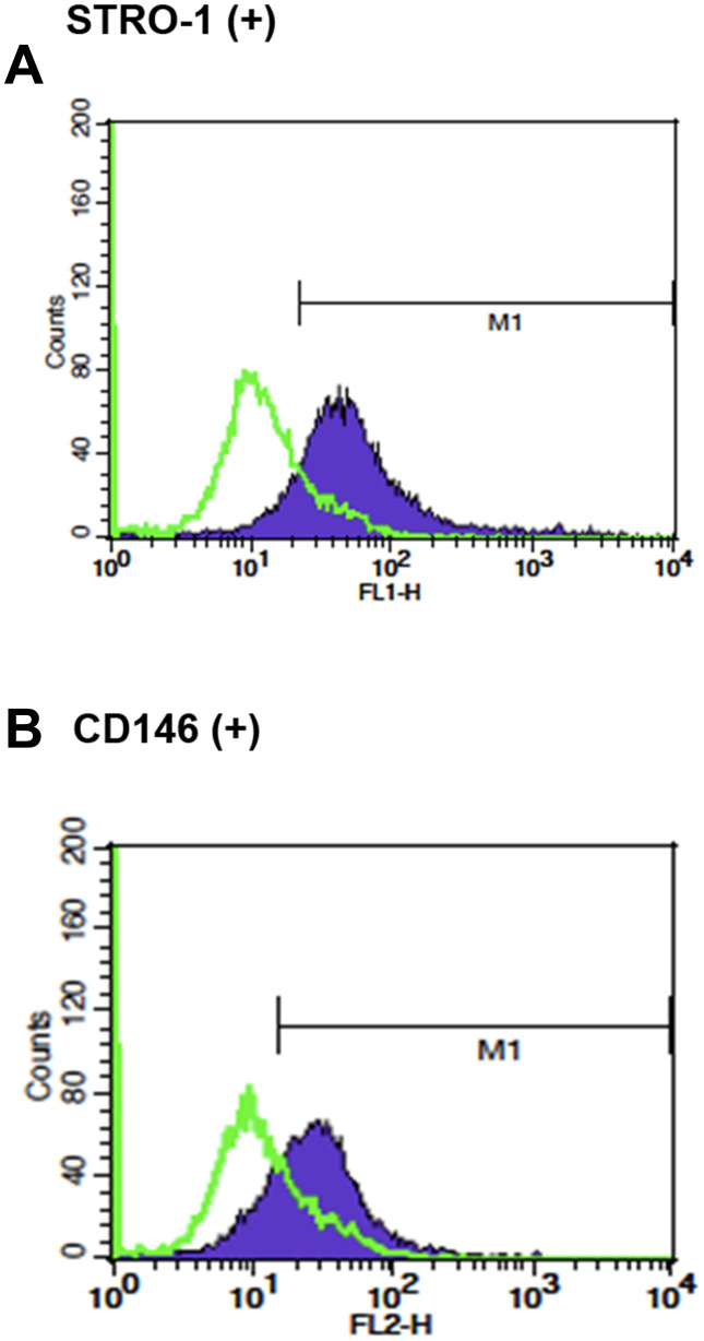
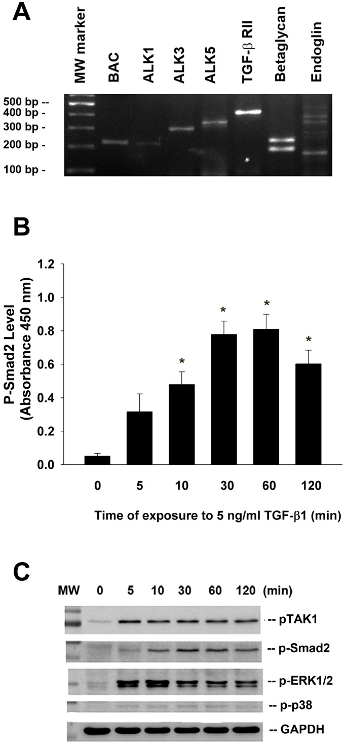
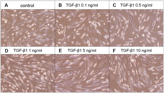
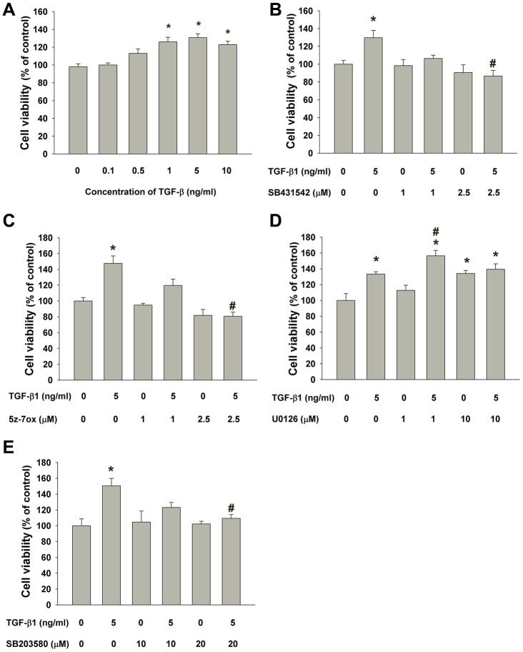
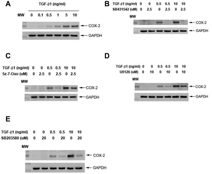
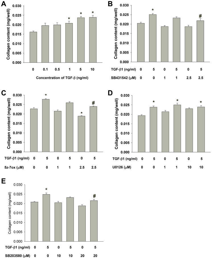
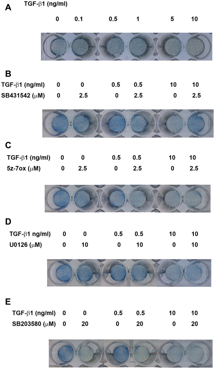
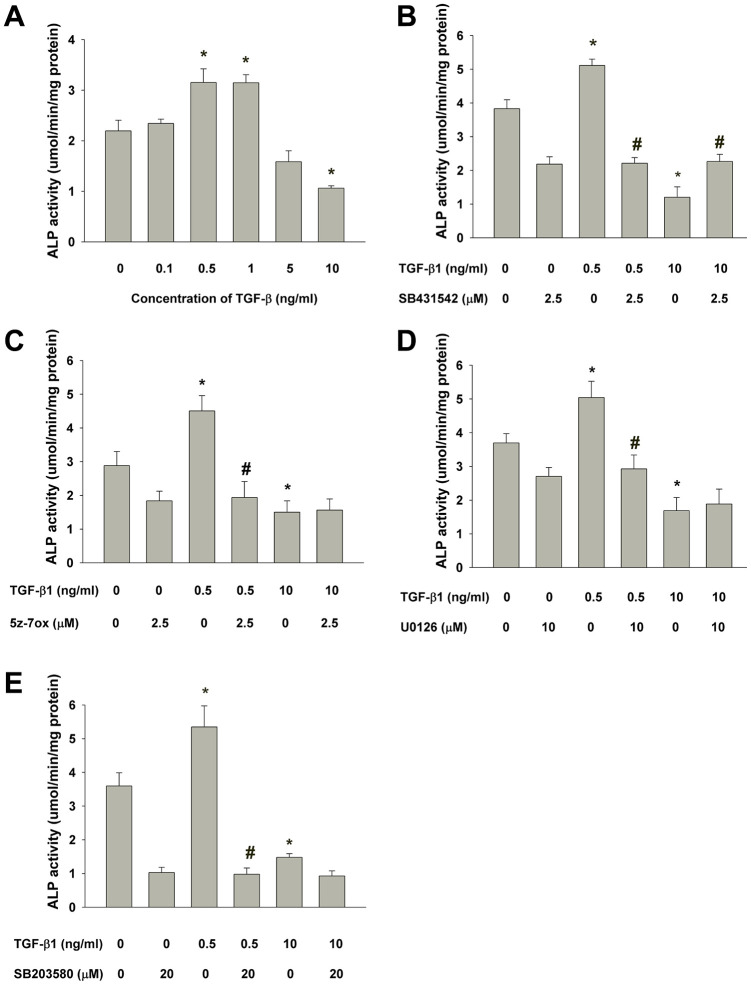
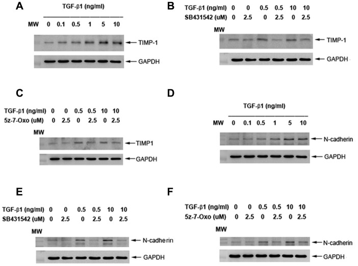
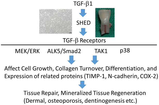
Transforming growth factor-β1 (TGF-β1) regulates wound healing/regeneration and aging processes. Dental pulp stem cells from human exfoliated deciduous teeth (SHED) are cell sources for treatment of age-related disorders. We studied the effect of TGF-β1 on SHED and related signaling. SHED were treated with TGF-β1 with/without pretreatment/co-incubation by SB431542, U0126, 5Z-7-oxozeaenol or SB203580. Sircol collagen assay, 3-(4,5-Dimethylthiazol-2-yl)-2,5- diphenyl tetrazolium bromide (MTT) assay, alkaline phosphatase (ALP) assay, RT-PCR, western blotting and PathScan phospho-ELISA were used to measure the effects. We found that SHED expressed ALK1, ALK3, ALK5, TGF-RII, betaglycan and endoglin mRNA. TGF-β1 stimulated p-Smad2, p-TAK1, p-ERK, p-p38 and cyclooxygenase-2 (COX-2) protein expression. It enhanced proliferation and collagen content of SHED that were attenuated by SB431542, 5Z-7-oxozeaenol and SB203580, but not U0126. TGF-β1 (0.5-1 ng/ml) stimulated ALP of SHED, whereas 5-10 ng/ml TGF-β1 suppressed ALP. SB431542 reversed the effects of TGF-β1. However, 5Z-7-oxozeaenol, SB203580 and U0126 only reversed the stimulatory effect of TGF-β1 on ALP. Four inhibitors attenuated TGF-β1-induced COX-2 expression. TGF-β1-stimulated TIMP-1 and N-cadherin was inhibited by SB431542 and 5Z-7-oxozeaenol. These results indicate that TGF-β1 affects SHED by differential regulation of ALK5/Smad2/3, TAK1, p38 and MEK/ERK. TGF-β1 and SHED could potentially be used for tissue engineering/regeneration and treatment of age-related diseases.
Keywords: aging-related diseases; alkaline phosphatase; collagen; cyclooxygenase-2; differentiation.
Conflict of interest statement
Figures
Similar articles
-
Effects of TGF-β1 on plasminogen activation in human dental pulp cells: Role of ALK5/Smad2, TAK1 and MEK/ERK signalling.J Tissue Eng Regen Med. 2018 Apr;12(4):854-863. doi: 10.1002/term.2339. Epub 2017 Apr 10. J Tissue Eng Regen Med. 2018. PMID: 27723266
-
Role of ALK5/Smad2/3 and MEK1/ERK Signaling in Transforming Growth Factor Beta 1-modulated Growth, Collagen Turnover, and Differentiation of Stem Cells from Apical Papilla of Human Tooth.J Endod. 2015 Aug;41(8):1272-80. doi: 10.1016/j.joen.2015.03.022. Epub 2015 May 19. J Endod. 2015. PMID: 26001858
-
TGF-β1 stimulates cyclooxygenase-2 expression and PGE2 production of human dental pulp cells: Role of ALK5/Smad2 and MEK/ERK signal transduction pathways.J Formos Med Assoc. 2017 Oct;116(10):748-754. doi: 10.1016/j.jfma.2017.07.008. Epub 2017 Aug 2. J Formos Med Assoc. 2017. PMID: 28779848
-
Regenerative Potential of Stem Cells Derived from Human Exfoliated Deciduous (SHED) Teeth during Engineering of Human Body Tissues.Curr Stem Cell Res Ther. 2021;16(5):507-517. doi: 10.2174/1574888X16999201231213206. Curr Stem Cell Res Ther. 2021. PMID: 33390148 Review.
-
[Dental pulp stem cells in tooth regeneration: advancement and emerging directions].Zhonghua Kou Qiang Yi Xue Za Zhi. 2024 May 9;59(5):496-501. doi: 10.3760/cma.j.cn112144-20240130-00048. Zhonghua Kou Qiang Yi Xue Za Zhi. 2024. PMID: 38637004 Review. Chinese.
Cited by
-
The growth factor multimodality on treating human dental mesenchymal stem cells: a systematic review.BMC Oral Health. 2024 Mar 1;24(1):290. doi: 10.1186/s12903-024-04013-2. BMC Oral Health. 2024. PMID: 38429689 Free PMC article.
-
Inducing cyclooxygenase-2 expression, prostaglandin E2 and prostaglandin F2α production of human dental pulp cells by activation of toll-like receptor-3, mitogen-activated protein kinase kinase/extracellular signal-regulated kinase and p38 signaling.J Dent Sci. 2024 Apr;19(2):1190-1199. doi: 10.1016/j.jds.2023.11.009. Epub 2023 Nov 25. J Dent Sci. 2024. PMID: 38618082 Free PMC article.
-
bFGF stimulated plasminogen activation factors, but inhibited alkaline phosphatase and SPARC in stem cells from apical Papilla: Involvement of MEK/ERK, TAK1 and p38 signaling.J Adv Res. 2022 Sep;40:95-107. doi: 10.1016/j.jare.2021.12.006. Epub 2021 Dec 28. J Adv Res. 2022. PMID: 36100336 Free PMC article.
-
Hopes and opportunities of stem cells from human exfoliated deciduous teeth (SHED) in cartilage tissue regeneration.Front Bioeng Biotechnol. 2023 Feb 13;11:1021024. doi: 10.3389/fbioe.2023.1021024. eCollection 2023. Front Bioeng Biotechnol. 2023. PMID: 36860887 Free PMC article. Review.
-
Bone morphogenetic protein 7 mediates stem cells migration and angiogenesis: therapeutic potential for endogenous pulp regeneration.Int J Oral Sci. 2022 Jul 20;14(1):38. doi: 10.1038/s41368-022-00188-y. Int J Oral Sci. 2022. PMID: 35858911 Free PMC article.
References
Publication types
MeSH terms
Substances
LinkOut - more resources
Full Text Sources
Medical
Research Materials
Miscellaneous
