

Direct drainage of the basal vein of Rosenthal into the superior petrosal sinus: a literature review
- PMID: 33148874
- PMCID: PMC7769095
- DOI: 10.5115/acb.20.199
Direct drainage of the basal vein of Rosenthal into the superior petrosal sinus: a literature review
Abstract
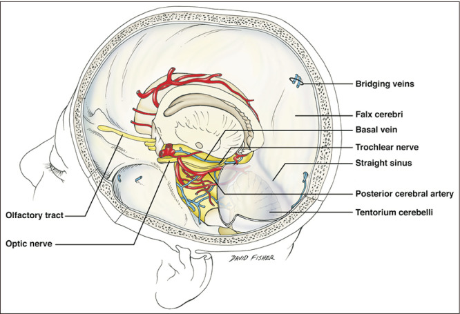
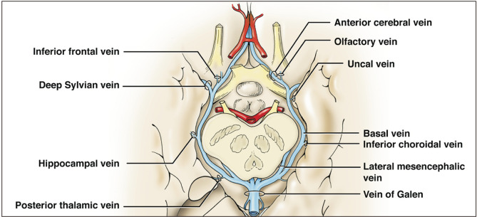
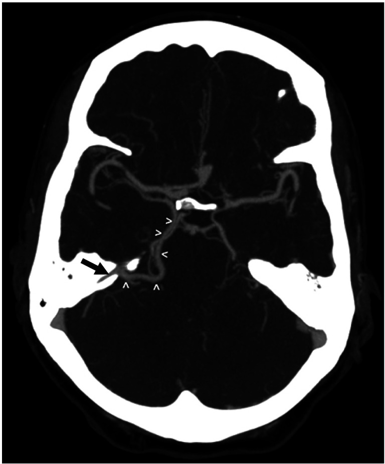
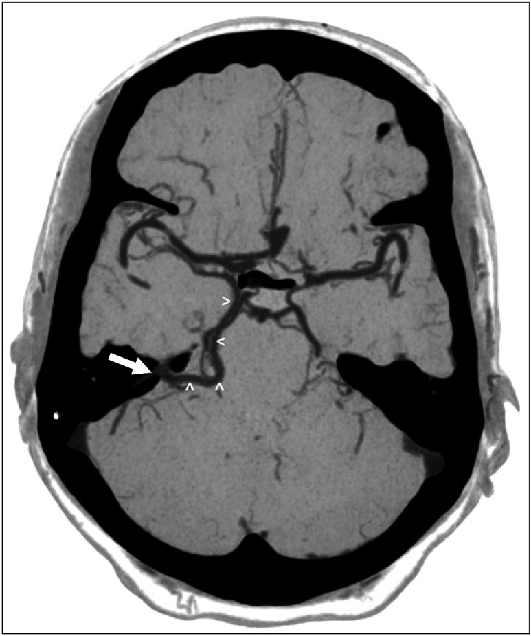
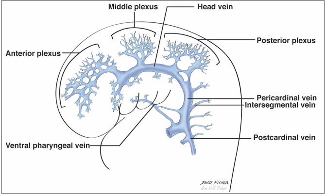
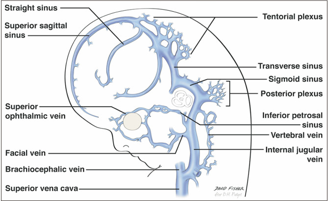
An adult male was found to have a variation of the left basal vein of Rosenthal after presenting with complaints of headache and balance issues. In this case, the vein drained directly into the left superior petrosal sinus (SPS) instead of the great vein of Galen. Anatomical variation of the basal vein is likely due to embryonic development of the deep cerebral venous system as primitive structures either differentiate regress or further with age. These changes may result in the uncommon presentation seen in this case. To our knowledge, this is the first case that shows the basal vein drains into the SPS. The normal and variant anatomy of this vessel are discussed.
Keywords: Basal vein; Basal vein of Rosenthal; Superior petrosal sinus; Tentorial sinus.
Conflict of interest statement
No potential conflict of interest relevant to this article was reported.
Figures
Similar articles
-
Drainage of the basal vein of Rosenthal into the confluence of sinuses.Anat Cell Biol. 2019 Jun;52(2):214-216. doi: 10.5115/acb.2019.52.2.214. Epub 2019 Jun 30. Anat Cell Biol. 2019. PMID: 31338241 Free PMC article.
-
Absence of the superior petrosal veins and sinus: Surgical considerations.Surg Neurol Int. 2015 Feb 26;6:34. doi: 10.4103/2152-7806.152147. eCollection 2015. Surg Neurol Int. 2015. PMID: 25745589 Free PMC article.
-
Anatomic variations of the deep cerebral veins,tributaries of Basal vein of rosenthal: embryologic aspects of the regressed embryonic tentorial sinus.Interv Neuroradiol. 2005 Jun 30;11(2):123-30. doi: 10.1177/159101990501100202. Epub 2005 Oct 25. Interv Neuroradiol. 2005. PMID: 20584491 Free PMC article.
-
Consequences of the anatomy of deep venous outflow from the brain.Neuroradiology. 1999 Apr;41(4):233-41. doi: 10.1007/s002340050739. Neuroradiology. 1999. PMID: 10344506 Review.
-
Spontaneous Resolution of Venous Aneurysms After Transarterial Embolization of a Variant Superior Sagittal Sinus Dural Arteriovenous Fistula: Case Report and Literature Review.Neurologist. 2017 Sep;22(5):186-195. doi: 10.1097/NRL.0000000000000137. Neurologist. 2017. PMID: 28859024 Review.
Cited by
-
Dural Arteriovenous Fistula Mimicking Acute Encephalitis.Intern Med. 2024 Feb 1;63(3):451-455. doi: 10.2169/internalmedicine.1819-23. Epub 2023 May 31. Intern Med. 2024. PMID: 37258162 Free PMC article. Review.
-
Combined Petrosal Intertentorial Approach: A Cadaveric Study of Comparison With the Standard Combined Petrosectomy.Oper Neurosurg. 2025 Jan 1;28(1):96-106. doi: 10.1227/ons.0000000000001244. Epub 2024 Jun 25. Oper Neurosurg. 2025. PMID: 38917345 Free PMC article.
-
Cadaveric findings of a duplicated superior petrosal sinus.Anat Cell Biol. 2022 Sep 30;55(3):384-389. doi: 10.5115/acb.22.012. Anat Cell Biol. 2022. PMID: 36168781 Free PMC article.
-
Morphometric evaluation of great vein of Galen and its clinical implications.Anat Cell Biol. 2023 Mar 31;56(1):32-38. doi: 10.5115/acb.22.051. Epub 2022 Oct 11. Anat Cell Biol. 2023. PMID: 36216783 Free PMC article.
-
Tentorial peeling during combined petrosal approach: a cadaveric dissection.Acta Neurochir (Wien). 2022 Nov;164(11):2833-2839. doi: 10.1007/s00701-022-05370-z. Epub 2022 Sep 26. Acta Neurochir (Wien). 2022. PMID: 36163381
References
-
- Huang YP, Wolf BS. The basal cerebral vein and its tributaries. In: Newton TH, Potts DG, editors. Radiology of the Skull and Brain, Vol. 2: Angiography. Mosby; Saint Louis: 1974. pp. 2111–54.
-
- Uddin MA, Haq TU, Rafique MZ. Cerebral venous system anatomy. J Pak Med Assoc. 2006;56:516–9. - PubMed
Publication types
LinkOut - more resources
Full Text Sources