. 2020 Nov 4;11(1):5577.
doi: 10.1038/s41467-020-19303-w.

Introducing Biomedisa as an open-source online platform for biomedical image segmentation

Affiliations

Introducing Biomedisa as an open-source online platform for biomedical image segmentation

Philipp D Lösel et al. Nat Commun. .

Erratum in

Abstract

We present Biomedisa, a free and easy-to-use open-source online platform developed for semi-automatic segmentation of large volumetric images. The segmentation is based on a smart interpolation of sparsely pre-segmented slices taking into account the complete underlying image data. Biomedisa is particularly valuable when little a priori knowledge is available, e.g. for the dense annotation of the training data for a deep neural network. The platform is accessible through a web browser and requires no complex and tedious configuration of software and model parameters, thus addressing the needs of scientists without substantial computational expertise. We demonstrate that Biomedisa can drastically reduce both the time and human effort required to segment large images. It achieves a significant improvement over the conventional approach of densely pre-segmented slices with subsequent morphological interpolation as well as compared to segmentation tools that also consider the underlying image data. Biomedisa can be used for different 3D imaging modalities and various biomedical applications.


Conflict of interest statement

The authors declare no competing interests.

Figures

Fig. 1
Fig. 1. Comparison between a conventional segmentation approach and Biomedisa.
Both procedures require manual pre-segmentation of the 3D image stack. While the widely used morphological interpolation solely considers labels on pre-segmented slices, Biomedisa takes both the underlying 3D image data and the pre-segmented slices into account, resulting in a significantly lower amount of required manual input. Moreover, interpolation artifacts are avoided and fine details like hairs, which are usually omitted during manual segmentation, are included.
Fig. 2
Fig. 2. Biomedisa segmentation using a Trigonopterus weevil as an example.
a Photograph of the original specimen. b Result of Biomedisa segmentation based on 37 pre-segmented slices of the tomographic volume adapted to the weevil’s morphology. c The 64 isolated body parts of (b). The surface meshes shown in this figure are based on the original Biomedisa result. If necessary, outliers or minor flaws in the reconstruction (e.g. tiny holes) can be corrected with low effort.
Fig. 3
Fig. 3. Biomedisa results based on different numbers of pre-segmented slices.
Inputs of 215 (as used for morphological interpolation), 108 and 54 equally spaced slices that correspond to pre-segmentation of every 5th, 10th and 20th slice provided accurate results. By adapting the spacing between the slices to the weevil’s morphology, a much lower count of only 37 slices yielded a dataset of equal quality. Lower numbers of pre-segmented slices resulted in increasingly flawed outputs.
Fig. 4
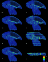
Fig. 4. Uncertainty of Biomedisa results based on different numbers of pre-segmented slices.
For 215, 108, 54 and 37 pre-segmented slices, the results show approximately the same degree of uncertainty, while for 27 or fewer slices the uncertainty increases significantly. The conspicuous bright line represents the boundary between elytra and thorax, which are closely interlocked. The boundary is almost invisible in the tomographic scan, thus resulting in a high uncertainty of the segmentation result. The uncertainty values range from 0 (blue) to 1 (red), with 0 meaning no uncertainty and 1 meaning a high degree of uncertainty, i.e. a voxel can be assigned to at least two labels with the same probability.
Fig. 5
Fig. 5. Biomedisa examples.
a Medaka fish with segmented skeleton and selected internal organs (based on µCT scan). b Mouse molar tooth showing enamel (white) and dentine (yellow) (µCT). c Human heart with segmented heart muscle and blood vessels (MRI). d Fossil parasitoid wasp from Baltic amber (SR-µCT). e Tracheal system of a hissing cockroach (µCT). f Claw of a theropod dinosaur from Burmese amber (SR-µCT). g Fossil parasitoid wasp preserved inside a mineralized fly pupa (SR-µCT). h Head of an Australian bull ant queen (SR-µCT). See “Methods” and Supplementary Table 2 for details on the specimens.
Fig. 6
Fig. 6. Speedup of computing times of GPU-based Biomedisa compared to CPU-based segmentation tools.
Speedup of computing times of different semi-automatic segmentation tools that take the image data into account compared to the slowest method according to Table 1. The values for mouse molar teeth and human hearts are average values.
Fig. 7
Fig. 7. Visual comparison of robustness to input errors.
Visual comparison of different segmentation methods for segmenting a human mandible based on flawed and inconsistent ground truth data labeled by two annotators. For the configuration, the values of the default parameters were chosen. If no default values were given, the values were selected from the examples in the documentation. Here, every 20th slice of the manual segmentation was used as initialization. The images show the segmentation result between two pre-segmented slices.

Similar articles

Cited by

References

    1. Maire, E. & Withers, P. J. Quantitative X-ray tomography. Int. Mater. Rev.59, 1–43 (2014).
    1. dos Santos Rolo, T., Ershov, A., van de Kamp, T. & Baumbach, T. In vivo X-ray cine-tomography for tracking morphological dynamics. Proc. Natl. Acad. Sci. USA111, 3921–3926 (2014). - PMC - PubMed
    1. Litjens, G. et al. A survey on deep learning in medical image analysis. Med. Image Anal.42, 60–88 (2017). - PubMed
    1. Christ, P. F. et al. Automatic liver and lesion segmentation in CT using cascaded fully convolutional neural networks and 3D conditional random fields. In Medical Image Computing and Computer-Assisted Intervention—MICCAI 2016 (eds Ourselin, S. et al.) 415–423 (Springer, Cham, 2016).
    1. Akkus, Z., Galimzianova, A., Hoogi, A., Rubin, D. L. & Erickson, B. J. Deep learning for brain MRI segmentation: state of the art and future directions. J. Digit. Imaging.30, 449–459 (2017). - PMC - PubMed

Publication types