. 2020 Nov 4;10(1):19065.
doi: 10.1038/s41598-020-76107-0.

Distinct effects of different matrix proteoglycans on collagen fibrillogenesis and cell-mediated collagen reorganization

Affiliations

Distinct effects of different matrix proteoglycans on collagen fibrillogenesis and cell-mediated collagen reorganization

Dongning Chen et al. Sci Rep. .

Abstract

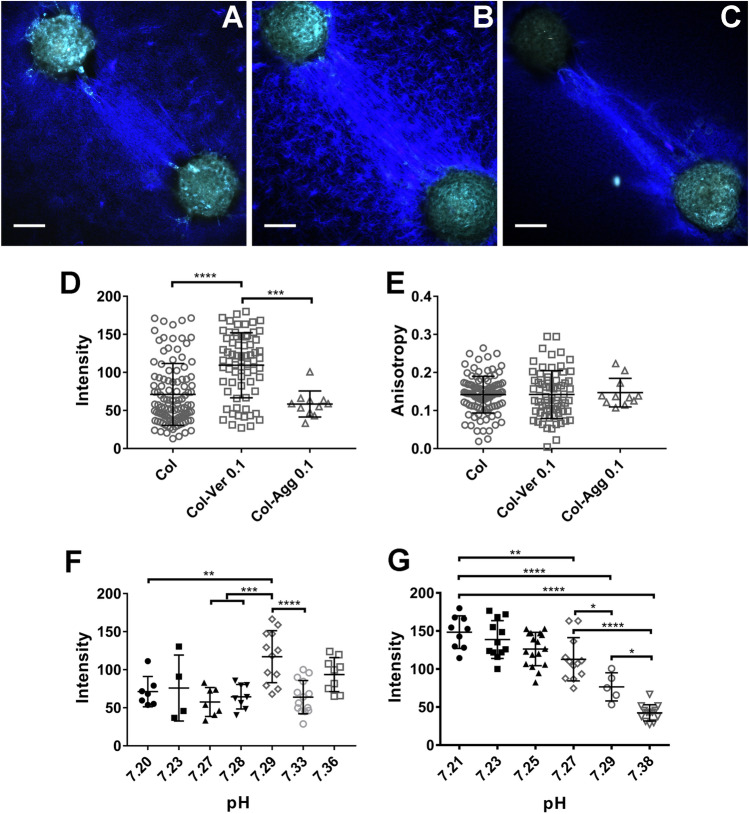
The extracellular matrix (ECM) is a complex mixture composed of fibrillar collagens as well as additional protein and carbohydrate components. Proteoglycans (PGs) contribute to the heterogeneity of the ECM and play an important role in its structure and function. While the small leucine rich proteoglycans (SLRPs), including decorin and lumican, have been studied extensively as mediators of collagen fibrillogenesis and organization, the function of large matrix PGs in collagen matrices is less well known. In this study, we showed that different matrix PGs have distinct roles in regulating collagen behaviors. We found that versican, a large chondroitin sulfate PG, promotes collagen fibrillogenesis in a turbidity assay and upregulates cell-mediated collagen compaction and reorganization, whereas aggrecan, a structurally-similar large PG, has different and often opposing effects on collagen. Compared to versican, decorin and lumican also have distinct functions in regulating collagen behaviors. The different ways in which matrix PGs interact with collagen have important implications for understanding the role of the ECM in diseases such as fibrosis and cancer, and suggest that matrix PGs are potential therapeutic targets.


Conflict of interest statement

The authors declare no competing interests.

Figures

Figure 1
Figure 1
Different matrix proteoglycans have distinct effects on collagen fibrillogenesis in an in vitro turbidity assay. (A) Versican (Ver; 0.1 mg/ml) was added to rat tail telocollagen (Col; 1.5 mg/ml) or to bovine atelocollagen (1.5 mg/ml). The experiments with telocollagen and atelocollagen were carried out at different times and have been combined to highlight the similar trend that occurs with the addition of versican. The assays were stopped after full gelation occurred, with a plateau in absorbance; this was significantly earlier for telo- than for atelocollagen. (B) Versican (Ver; purple curve) or aggrecan (Agg; blue curve), both at 0.1 mg/ml, were added to atelocollagen (Col; 1.5 mg/ml, black curve). Versican accelerated gelation dramatically while aggrecan slightly right-shifted the turbidity curve. (C) Versican alone (0.1 mg/ml) failed to gel and showed no change in turbidity over time under the assay conditions. (D) The SLRPs lumican (Lum; 0.01, 0.05 and 0.1 mg/ml) and decorin (Dec, 0.1 mg/ml) were added to atelocollagen (Col; 1.5 mg/ml). Decorin had a larger impact on decreasing fibrillogenesis than lumican. For all turbidity assays under all testing conditions, the pH and gelation temperature were the same. For all panels except (C), three independent experiments were carried out for each condition, each with three technical replicates. Because there can be day-to-day differences in the absolute absorbance values for the assay, a representative figure from one experiment with mean curves is shown for each condition; however, all assays in a panel were carried out in parallel, and relative values among the different conditions were consistent in each individual experiment. The dotted lines represent Standard Deviation (SD). (C) was performed once with three technical replicates; the dotted lines represent SD.
Figure 2
Figure 2
Versican core protein, with a minor contribution from the CS side chains, regulates collagen gelation. (A) Chondroitin sulfate (CS; 0.01, 0.04, 0.07 and 0.1 mg/ml; green, yellow, orange and red curves) was added to collagen (Col; 1.5 mg/ml; black curve). (B) Recombinant V3 isoform (V3, 0.1 mg/ml) was added to rat rail telocollagen (1.5 mg/ml) and bovine atelocollagen (1.5 mg/ml). This experiment was carried out as in Fig. 1A. (C) After digestion of the versican CS with ChABC, the remaining versican core protein was added at 0.1 mg/ml (pink curve) to atelocollagen (1.5 mg/ml; red curve) and caused a similar although slightly blunted right shift to the curves. Heat-inactivated ChABC had minimal effect on collagen gelation (blue curve). Three independent experiments were carried out for each condition, each with three technical replicates. Because there can be day-to-day differences in the absolute absorbance values for the assay, a representative figure from one experiment with mean curves is shown for each condition; however, all assays in a panel were carried out in parallel, and relative values among the different conditions were consistent in each individual experiment. The dotted lines represent SD.
Figure 3
Figure 3
Matrix PGs have different effects on the structure of collagen networks. (AE) Representative SEM images of telocollagen matrices with different PGs added. (A) Telocollagen (1.5 mg/ml) alone; (BE) Telocollagen (Col; 1.5 mg/ml) with 0.1 mg/ml versican (Ver) (B), 0.1 mg/ml aggrecan (Agg) (C), 0.05 mg/ml lumican (Lum) (D) and 0.1 mg/ml decorin (Dec) (E). (F,G) Quantification of fiber diameter and porosity using DiameterJ. Three independent experiments were carried out and one gel was generated for each condition in each experiment. 5 SEM images were taken for each gel at random locations. When analyzed using FibrilTool, 5 sections were cropped from each SEM image and a measurement was taken on each cropped figure. Each data point represents a single measurement. Scale bar = 1 µm. Data represent mean ± SD. ****P < 0.0001.
Figure 4
Figure 4
Large CS proteoglycans have differential effects on cell-mediated collagen reorganization. (AC) Representative SHG images of aligned collagen fibers between pairs of NIH 3T3 spheroids. Blue represents the SHG signal from collagen; green is cell autofluorescence. (A) collagen (Col; 1.5 mg/ml) alone, (B) collagen-versican (Ver; 0.1 mg/ml) and (C) collagen-aggrecan (Agg; 0.1 mg/ml) plugs. (DE) Intensity and anisotropy in the aligned collagen area for (AC). (F,G) Collagen compaction in pure collagen plugs (F) was not pH sensitive, but the impact of versican on collagen compaction was highly pH-dependent (G). Each data point in (DG) represents collagen behavior between one pair of spheroids. At least 3 independent experiments were carried out for each condition, with at least 3 pairs of plugs examined for each experiment. For the pH testing in (F and G), 4–12 pairs of spheroids were analyzed for each pH. Spheroids were seeded approximately 500 µm apart. Scale bars = 100 µm. Data represent mean ± SD. *P < 0.05, **P < 0.01, ***P < 0.001 and ****P < 0.0001.
Figure 5
Figure 5
SLRPs regulate cell-mediated collagen reorganization differently. (AD) Representative SHG images of collagen fibers between portal fibroblast spheroids on (A) collagen (1.5 mg/ml) alone, (B) collagen-decorin (Dec; 0.1 mg/ml) and (C,D) collagen-lumican (Lum, 0.01 or 0.05 mg/ml) plugs. (E,F) Quantification of cell-mediated collagen alignment with the addition of decorin and lumican, from (AD). Each data point in (E and F) represents collagen behavior between one pair of spheroids. At least 3 independent experiments were carried out for each condition, with at least 3 pairs of plugs in each experiment. Spheroids are seeded approximately 500 µm apart. Scale bar = 100 µm. Data represent mean ± SD. **P < 0.01, ***P < 0.001 and ****P < 0.0001.
Figure 6
Figure 6
Matrix PGs have different effects on the contraction of engineered collagen microtissues. (A,B) Representative light microscopy images of PDMS cantilever displacement in µTUGs. (C) SHG imaging of µTUGs made using collagen and NIH 3T3 fibroblasts. (D) Quantification of increased displacement observed with inclusion of 0.1 mg/ml versican (Ver) in 1.5 mg/ml collagen (Col) microtissue. (E) Quantification of the displacement observed in collagen microtissues with or without aggrecan (Agg; 0.1 mg/ml), decorin (Dec; 0.1 mg/ml), or lumican (Lum; 0.01 mg/ml or 0.05 mg/ml). N > 30 microtissues per each platform, three independent experiments (platforms) per condition. Points represent mean per platform. Scale bar = 200 µm. Data represent mean ± SE. *P < 0.05 and **P < 0.01.

Similar articles

Cited by

References

    1. Urbanczyk M, Layland SL, Schenke-Layland K. The role of extracellular matrix in biomechanics and its impact on bioengineering of cells and 3D tissues. Matrix Biol. 2020;85–86:1–14. doi: 10.1016/j.matbio.2019.11.005. - DOI - PubMed
    1. Abhilash AS, Baker BM, Trappmann B, Chen CS, Shenoy VB. Remodeling of fibrous extracellular matrices by contractile cells: Predictions from discrete fiber network simulations. Biophys. J. 2014;107:1829–1840. doi: 10.1016/j.bpj.2014.08.029. - DOI - PMC - PubMed
    1. Kalamajski S, Oldberg Å. The role of small leucine-rich proteoglycans in collagen fibrillogenesis. Matrix Biol. 2010;29:248–253. doi: 10.1016/j.matbio.2010.01.001. - DOI - PubMed
    1. Karamanos NK, Theocharis AD, Neill T, Iozzo RV. Matrix modeling and remodeling: A biological interplay regulating tissue homeostasis and diseases. Matrix Biol. 2019;75–76:1–11. doi: 10.1016/j.matbio.2018.08.007. - DOI - PMC - PubMed
    1. Yanagishita M. Function of proteoglycans in the extracellular matrix. Kokubyo Gakkai Zasshi. 1997;64:193–204. doi: 10.5357/koubyou.64.193. - DOI - PubMed

Publication types