. 2020 Nov 4;10(1):18974.
doi: 10.1038/s41598-020-75526-3.

Characterisation of the main PSA glycoforms in aggressive prostate cancer

Affiliations

Characterisation of the main PSA glycoforms in aggressive prostate cancer

Anna Gratacós-Mulleras et al. Sci Rep. .

Abstract

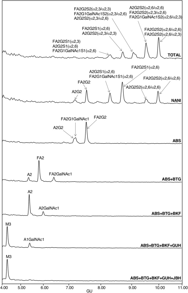
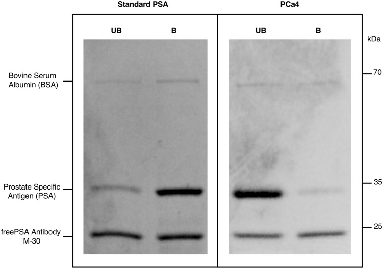
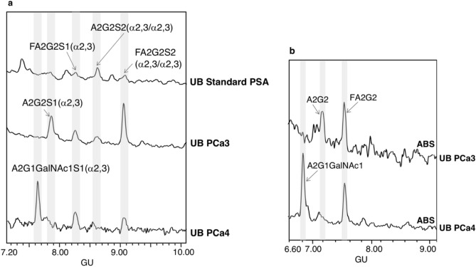
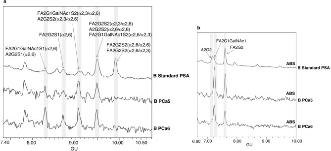
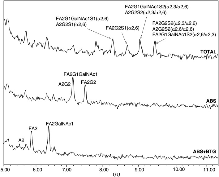
Serum levels of prostate specific antigen (PSA) are commonly used for prostate cancer (PCa) detection. However, their lack of specificity to distinguish benign prostate pathologies from PCa, or indolent from aggressive PCa have prompted the study of new non-invasive PCa biomarkers. Aberrant glycosylation is involved in neoplastic progression and specific changes in PSA glycosylation pattern, as the reduction in the percentage of α2,6-sialic acid (SA) are associated with PCa aggressiveness. In this study, we have characterised the main sialylated PSA glycoforms from blood serum of aggressive PCa patients and have compared with those of standard PSA from healthy individuals' seminal plasma. PSA was immunoprecipitated and α2,6-SA were separated from α2,3-SA glycoforms using SNA affinity chromatography. PSA N-glycans were released, labelled and analysed by hydrophilic interaction liquid chromatography combined with exoglycosidase digestions. The results showed that blood serum PSA sialylated glycoforms containing GalNAc residues were largely increased in aggressive PCa patients, whereas the disialylated core fucosylated biantennary structures with α2,6-SA, which are the major PSA glycoforms in standard PSA from healthy individuals, were markedly reduced in aggressive PCa. The identification of these main PSA glycoforms altered in aggressive PCa opens the way to design specific strategies to target them, which will be useful to improve PCa risk stratification.


Conflict of interest statement

The authors declare no competing interests.

Figures

Figure 1
Figure 1
HILIC-UPLC profiles of standard PSA N-glycans labelled with 2AB. From top to bottom, chromatograms of consecutive panels correspond to the total profile and the corresponding digestions by the specified exoglycosidases. Profiles are standardised against a dextran hydrolysate with glucose units (GU). Abbreviations used for the different structures are defined in Table 1.
Figure 2
Figure 2
Representative gel electrophoresis of fPSA immunoprecipitated from unbound (UB) and bound (B) fractions of SNA affinity chromatography. Results of standard PSA and PSA from prostate cancer (PCa4) corresponding to two different gels are shown.
Figure 3
Figure 3
HILIC-UPLC profiles of PSA N-glycans from unbound (UB) fractions. (a) Total UB fractions of standard PSA (top panel) and PSA from aggressive prostate cancer (PCa3-PCa4) (middle and bottom panel) and (b) ABS digested N-glycans from PSA from UB fractions of PCa3 (top panel) and PCa4 (bottom panel). Profiles are standardised against a dextran hydrolysate with glucose units (GU).
Figure 4
Figure 4
HILIC-UPLC profiles of PSA N-glycans from bound (B) fractions. (a) Total B fractions of standard PSA (top panel) and PSA from aggressive prostate cancer (PCa5-PCa6) (middle and bottom panel) and (b) ABS digested N-glycans of B fraction from standard PSA (top panel), PCa5 and PCa6 (middle and bottom panel). Profiles are standardised against a dextran hydrolysate with glucose units (GU).
Figure 5
Figure 5
HILIC-UPLC profiles of PSA N-glycans from aggressive prostate cancer (PCa6) bound (B) fraction: total profile (top panel), after ABS digestion (middle panel) and after ABS + BTG digestion (bottom panel). Profiles are standardised against a dextran hydrolysate with glucose units (GU).

Similar articles

Cited by

References

    1. Siegel RL, Miller KD, Jemal A. Cancer statistics, 2020. CA Cancer J. Clin. 2020;70:7–30. doi: 10.3322/caac.21590. - DOI - PubMed
    1. Schroder FH, et al. Evaluation of the digital rectal examination as a screening test for prostate cancer. J. Natl. Cancer Inst. 1998;90:1817–1823. doi: 10.1093/jnci/90.23.1761. - DOI - PubMed
    1. Lilja H, Ulmert D, Vickers AJ. Prostate-specific antigen and prostate cancer: prediction, detection and monitoring. Nat. Rev. Cancer. 2008;8:268–278. doi: 10.1038/nrc2351. - DOI - PubMed
    1. Fuessel S, Wirth MP. New markers in prostate cancer: genomics. Arch. Esp. Urol. 2019;72:116–125. - PubMed
    1. Crawford ED, Denes BS, Ventii KH, Shore ND. Prostate cancer: incorporating genomic biomarkers in prostate cancer decisions. Clin. Pract. 2014;11:605–612. doi: 10.2217/cpr.14.65. - DOI

Publication types