1, 6-O, O-Diacetylbritannilactone from Inula britannica Induces Anti-Tumor Effect on Oral Squamous Cell Carcinoma via miR-1247-3p/LXRα/ABCA1 Signaling
- PMID: 33149621
- PMCID: PMC7605651
- DOI: 10.2147/OTT.S263014
1, 6-O, O-Diacetylbritannilactone from Inula britannica Induces Anti-Tumor Effect on Oral Squamous Cell Carcinoma via miR-1247-3p/LXRα/ABCA1 Signaling
Abstract
Introduction: Oral squamous cell carcinoma (OSCC) is the most prevalent malignancy affecting the oral cavity and is associated with severe morbidity and high mortality. 1, 6-O, O-Diacetylbritannilactone (OODBL) isolated from the medicinal herb of Inula britannica has various biological activities such as anti-inflammation and anti-cancer. However, the effect of OODBL on OSCC progression remains unclear. Here, we were interested in the function of OODBL in the development of OSCC.
Methods: The effect of OODBL on OSCC progression was analyzed by MTT assays, colony formation assays, transwell assays, apoptosis analysis, cell cycle analysis, and in vivo tumorigenicity analysis. The mechanism investigation was performed by qPCR assays, Western blot analysis, and luciferase reporter gene assays.
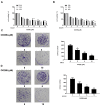
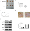
Results: We found that OODBL inhibits the proliferation of OSCC cells in vitro. Moreover, the migration and invasion were attenuated by OODBL treatment in the OSCC cells. OODBL arrested cells at the G0/G1 phase and induced cell apoptosis. OODBL was able to up-regulate the expression of LXRα, ABCA1, and ABCG1 in the system. In addition, OODBL activated LXRα/ABCA1 signaling by targeting miR-1247-3p. Furthermore, the expression levels of cytochrome c in the cytoplasm, cleaved caspase-9, and cleaved caspase-3 were dose-dependently reduced by OODBL. Besides, OODBL increased the expression ratio of Bax to Bcl-2. Moreover, OODBL repressed tumor growth of OSCC cells in vivo.
Discussion: Thus, we conclude that OODBL inhibits OSCC progression by modulating miR-1247-3p/LXRα/ABCA1 signaling. Our finding provides new insights into the mechanism by which OODBL exerts potent anti-tumor activity against OSCC. OODBL may be a potential anti-tumor candidate, providing a novel clinical treatment strategy of OSCC.
Keywords: LXRα/ABCA1 signaling; OODBL; OSCC; anti-tumor; miR-1247-3p.
© 2020 Zheng et al.
Conflict of interest statement
The authors declare no competing financial or non-financial interests. Shaohua Zheng and Lihua Li contributed equally to this work.
Figures


Similar articles
-
Involvement of MAPK, Bcl-2 family, cytochrome c, and caspases in induction of apoptosis by 1,6-O,O-diacetylbritannilactone in human leukemia cells.Mol Nutr Food Res. 2007 Feb;51(2):229-38. doi: 10.1002/mnfr.200600148. Mol Nutr Food Res. 2007. PMID: 17262884
-
MiR-376c-3p regulates the proliferation, invasion, migration, cell cycle and apoptosis of human oral squamous cancer cells by suppressing HOXB7.Biomed Pharmacother. 2017 Jul;91:517-525. doi: 10.1016/j.biopha.2017.04.050. Epub 2017 May 5. Biomed Pharmacother. 2017. PMID: 28482289
-
CircKRT1 drives tumor progression and immune evasion in oral squamous cell carcinoma by sponging miR-495-3p to regulate PDL1 expression.Cell Biol Int. 2021 Jul;45(7):1423-1435. doi: 10.1002/cbin.11581. Epub 2021 Apr 14. Cell Biol Int. 2021. PMID: 33675276
-
1,6-O,O-Diacetylbritannilactone Inhibits Eotaxin-1 and ALOX15 Expression Through Inactivation of STAT6 in A549 Cells.Inflammation. 2017 Dec;40(6):1967-1974. doi: 10.1007/s10753-017-0637-y. Inflammation. 2017. PMID: 28770377
-
MicroRNA-141-3p inhibits the progression of oral squamous cell carcinoma via targeting PBX1 through the JAK2/STAT3 pathway.Exp Ther Med. 2022 Jan;23(1):97. doi: 10.3892/etm.2021.11020. Epub 2021 Dec 1. Exp Ther Med. 2022. PMID: 34976139 Free PMC article.
Cited by
-
Emerging Insights into Liver X Receptor α in the Tumorigenesis and Therapeutics of Human Cancers.Biomolecules. 2023 Jul 28;13(8):1184. doi: 10.3390/biom13081184. Biomolecules. 2023. PMID: 37627249 Free PMC article. Review.
-
Strategies to gain novel Alzheimer's disease diagnostics and therapeutics using modulators of ABCA transporters.Free Neuropathol. 2021;2:33. doi: 10.17879/freeneuropathology-2021-3528. Epub 2021 Dec 13. Free Neuropathol. 2021. PMID: 34977908 Free PMC article.
-
Tumor-derived exosomal miR-1247-3p promotes angiogenesis in bladder cancer by targeting FOXO1.Cancer Biol Ther. 2024 Dec 31;25(1):2290033. doi: 10.1080/15384047.2023.2290033. Epub 2023 Dec 10. Cancer Biol Ther. 2024. PMID: 38073044 Free PMC article.
-
miR-1247-3p regulation of CCND1 affects chemoresistance in colorectal cancer.PLoS One. 2024 Dec 31;19(12):e0309979. doi: 10.1371/journal.pone.0309979. eCollection 2024. PLoS One. 2024. PMID: 39739897 Free PMC article.
-
miR-1247-3p targets STAT5A to inhibit lung adenocarcinoma cell migration and chemotherapy resistance.J Cancer. 2022 Mar 28;13(7):2040-2049. doi: 10.7150/jca.65167. eCollection 2022. J Cancer. 2022. PMID: 35517418 Free PMC article.
References
LinkOut - more resources
Full Text Sources
Research Materials

