. 2020 Oct 16:2020:4828256.
doi: 10.1155/2020/4828256. eCollection 2020.

MUTYH Deficiency Is Associated with Attenuated Pulmonary Fibrosis in a Bleomycin-Induced Model

Affiliations

MUTYH Deficiency Is Associated with Attenuated Pulmonary Fibrosis in a Bleomycin-Induced Model

Qingmin Sun et al. Oxid Med Cell Longev. .

Abstract

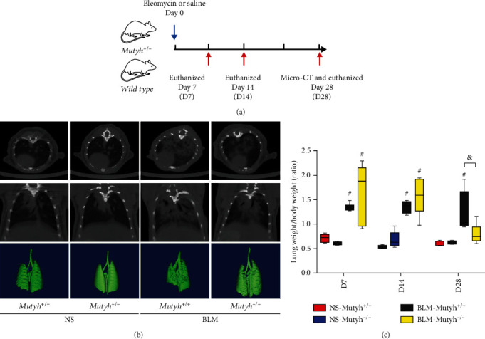
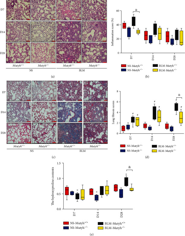
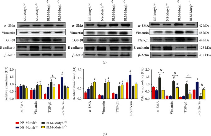
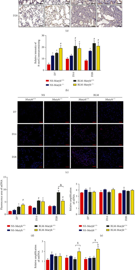
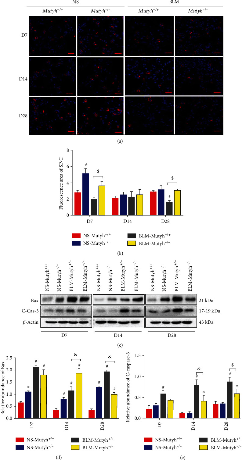
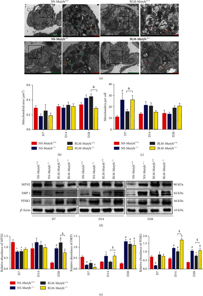
Idiopathic pulmonary fibrosis (IPF) is a progressive, irreversible lung disease of unknown etiology with limited survival. IPF incidence and prevalence increase significantly with aging, which is associated with an age-related accumulation of oxidative DNA damage. The Mutyh gene is involved in the base excision repair (BER) system, which is critical for repairing the misincorporated adenine that is opposite to the oxidized guanine base, 8-oxoguanine, and maintaining the fidelity of DNA replication. We used Mutyh knockout mice and a bleomycin-induced pulmonary fibrosis model to test the effect of MUTYH deficiency on lesion progression. Unexpectedly, a much less severe lesion of pulmonary fibrosis was observed in Mutyh -/- than in Mutyh +/+ mice, which was supported by assay on protein levels of TGF-β1 and both fibrotic markers, α-SMA and Vimentin, in pulmonary tissues of the model animals. Mechanically, MUTYH deficiency prevented the genomic DNA of pulmonary tissue cells from the buildup of single-strand breaks (SSBs) of DNA and maintained the integrity of mtDNA. Furthermore, increased mitochondrial dynamic regulation and mitophagy were detected in pulmonary tissues of the bleomycin-induced Mutyh -/- model mice, which could reduce the pulmonary epithelial cell apoptosis. Our results suggested that MUTYH deficiency could even induce protective responses of pulmonary tissue under severe oxidative stress.


Conflict of interest statement

The authors declare no conflicts of interest.

Figures

Figure 1
Figure 1
The bleomycin- (BLM-) induced pulmonary fibrosis mouse model was evaluated using Micro-CT and lung weight/body weight ratios. (a) Schematic representation of the experimental design is shown. (b) Micro-CT and 3D digital reconstruction were employed to directly observe the fibrotic lesions in the lungs of mice on day 28 after intratracheal administration with a single dose of BLM or normal saline (NS) on day 0. (c) Lung weight/body weight ratios are shown compared to NS groups. BLM treatment markedly increased the ratios at all three time points. On day 28, BLM-Mutyh−/− mice exhibited a significantly lower ratio of lung weight/body weight, compared to BLM-Mutyh+/+ mice (n ≥ 5, P = 0.006). Statistical significance was analyzed using two-way ANOVA and followed by the LSD post hoc test. ∗ represents P < 0.05 and # represents P < 0.01 compared with the NS-Mutyh+/+ group. & represents P < 0.01 compared to the BLM-Mutyh+/+ group.
Figure 2
Figure 2
MUTYH-deficient mice exhibited attenuated pulmonary fibrosis in the BLM-induced model. (a) Representative histological sections of pulmonary tissues were obtained from experimental animals and subjected to HE staining. (b) The inflammatory score based on HE staining in the BLM-Mutyh−/− group was significantly lower than that in BLM-Mutyh+/+ among D7 mice (P = 0.003). (c, d) Representative images of Masson's trichrome staining in pulmonary tissue sections revealed significantly decreased collagen deposition (blue) in BLM-Mutyh−/− compared to BLM-Mutyh+/+ mice among the D28 group (P < 0.001). Scale bars: 100 μm. (e) Collagen deposition was assessed by hydroxyproline contents. BLM-Mutyh−/− exhibited significantly lower contents compared to BLM-Mutyh+/+ mice among the D28 group (P = 0.004). n ≥ 5 mice per group. Statistical significance was analyzed using two-way ANOVA and followed by the LSD post hoc test. ∗ represents P < 0.05 and # represents P < 0.01 compared with NS-Mutyh+/+ mice. & represents P < 0.01 compared to the BLM-Mutyh+/+ group.
Figure 3
Figure 3
Mutyh −/−-associated reduction of TGF-β1 and fibrotic marker expression in BLM-induced model mice. (a) Western blot showed an increased expression of TGF-β1 and fibrotic markers in pulmonary tissues from BLM-treated mice, especially in D14 and D28. In D28, MUTYH deficiency significantly reduced the levels of α-SMA, Vimentin, and TGF-β1 compared to BLM-Mutyh+/+ mice, while an upregulation expression of E-cadherin was observed in NS-Mutyh−/− mice of all three groups and in BLM-Mutyh−/− of D28. (b) Histograms of the protein measure show the relative expression of each protein (n = 3). Statistical significance was analyzed using two-way ANOVA and followed by the LSD post hoc test. ∗ represents P < 0.05 and # represents P < 0.01 compared to the NS-Mutyh+/+ group. $ represents P < 0.05 compared to the BLM-Mutyh+/+ group. $ represents P < 0.05 and & represents P < 0.01 compared to the BLM-Mutyh+/+ group.
Figure 4
Figure 4
MUTYH deficiency alleviated SSB formation and maintained mtDNA integrity. (a, b) Representative images of immunostaining of 8-oxoG and quantification of the intensity of 8-oxoG immunostaining in pulmonary tissue sections (n = 4 for per genotype/treatment; scale bar: 100 μm). (c, d) Pulmonary tissue specimens of mice were subjected to immunofluorescence staining with anti-ssDNA (red, original magnification: ×630, n = 4, and scale bar: 67 μm). The immune complexes and DAPI-stained nuclei (blue) were visualized under a fluorescence microscope. Total fluorescence intensity of 4 fields selected randomly in 3 sections was quantified with ImageJ (b). (e, f) An 8.7 kb fragment from β-globin loci and a 10 kb fragment from mtDNA were used for measuring nDNA and mtDNA integrity, respectively. At least two independent experiments were performed for each sample (n ≥ 5). Statistical significance was analyzed using two-way ANOVA and followed by the LSD post hoc test. ∗ represents P < 0.05 and # represents P < 0.01 compared with the NS-Mutyh+/+ group. $ represents P < 0.05 compared to the BLM-Mutyh+/+ group. $ represents P < 0.05 and & represents P < 0.01 compared to the BLM-Mutyh+/+ group.
Figure 5
Figure 5
Apoptosis of epithelial cells was suppressed in Mutyh−/− mice treated with BLM. (a) Pulmonary tissue sections of mice were subjected to immunofluorescence staining with anti-SP-C (red, original magnification: ×630, n = 4, and scale bar: 67 μm). Merged images with DAPI-stained nuclei were shown. (b) Total fluorescence intensity of SP-C was quantified with ImageJ. (c) Protein extracts from pulmonary tissues of different groups were subjected to Western blotting with anti-Bax and Cleaved-caspase-3. (d, e) Histograms of the protein measure show the relative expression of each protein (n = 3). Statistical significance was analyzed using two-way ANOVA and followed by the LSD post hoc test. ∗ represents P < 0.05 and # represents P < 0.01 compared with the NS-Mutyh+/+ group. $ represents P < 0.05 and & represents P < 0.01 compared to the BLM-Mutyh+/+ group.
Figure 6
Figure 6
MUTYH deficiency maintained mitochondrial homeostasis. (a) Representative images of transmission electron microscopy (TEM) of lung AECIIs from different groups of mice (lamellar bodies used as identification marks, orange arrows; green scale bar: 5 μm, n = 4). The boxed region of the image is magnified in the right panel (red scale bar: 1 μm). Mitochondria in AECIIs of pulmonary tissues from model mice were mostly enlarged. The normal mitochondria were indicated with green arrows and dysmorphic ones with red arrows. (b) Quantitative morphometric analyses of the mitochondrial area in single AECII with TEM images. (c) Mitochondrial number per AECII was counted in the TEM images from different groups of mice. (d) Expression levels of the proteins involved in mitochondrial fusion/fission dynamic regulation. Western blotting with anti-MFN2, DRP1, and PINK1 was shown. (e) Histograms show the relative expression levels of MFN2, DRP1, and PINK1 proteins (n = 3). Statistical significance was analyzed using two-way ANOVA and followed by the LSD post hoc test. ∗ represents P < 0.05 and # represents P < 0.01 compared with the NS-Mutyh+/+ group. $ represents P < 0.05 and & represents P < 0.01 compared to the BLM-Mutyh+/+ group.

Similar articles

Cited by

References

    1. King T. E., Jr., Pardo A., Selman M. Idiopathic pulmonary fibrosis. The Lancet. 2011;378(9807):1949–1961. doi: 10.1016/S0140-6736(11)60052-4. - DOI - PubMed
    1. Yue L., Yao H. Mitochondrial dysfunction in inflammatory responses and cellular senescence: pathogenesis and pharmacological targets for chronic lung diseases. British Journal of Pharmacology. 2016;173(15):2305–2318. doi: 10.1111/bph.13518. - DOI - PMC - PubMed
    1. Ayala-Peña S. Role of oxidative DNA damage in mitochondrial dysfunction and Huntington’s disease pathogenesis. Free Radical Biology and Medicine. 2013;62:102–110. doi: 10.1016/j.freeradbiomed.2013.04.017. - DOI - PMC - PubMed
    1. Valavanidis A., Vlachogianni T., Fiotakis C. 8-Hydroxy-2'-deoxyguanosine (8-OHdG): a critical biomarker of oxidative stress and carcinogenesis. Journal of environmental science and health Part C. 2009;27(2):120–139. doi: 10.1080/10590500902885684. - DOI - PubMed
    1. Al-Tassan N., Chmiel N. H., Maynard J., et al. Inherited variants of MYH associated with somatic G: C→ T: A mutations in colorectal tumors. Nature Genetics. 2002;30(2):227–232. doi: 10.1038/ng828. - DOI - PubMed

MeSH terms