

Infected Right Ventricle Thrombus as a Cause of Persistent Sepsis
- PMID: 33150103
- PMCID: PMC7603886
- DOI: 10.7759/cureus.10751
Infected Right Ventricle Thrombus as a Cause of Persistent Sepsis
Abstract
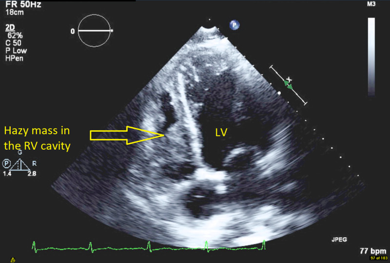
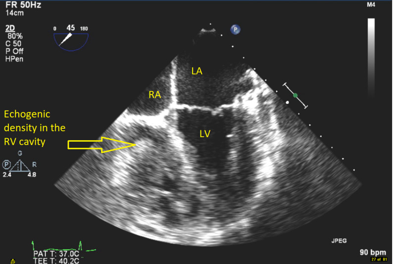
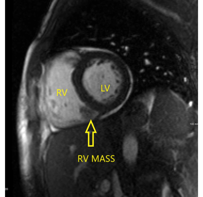
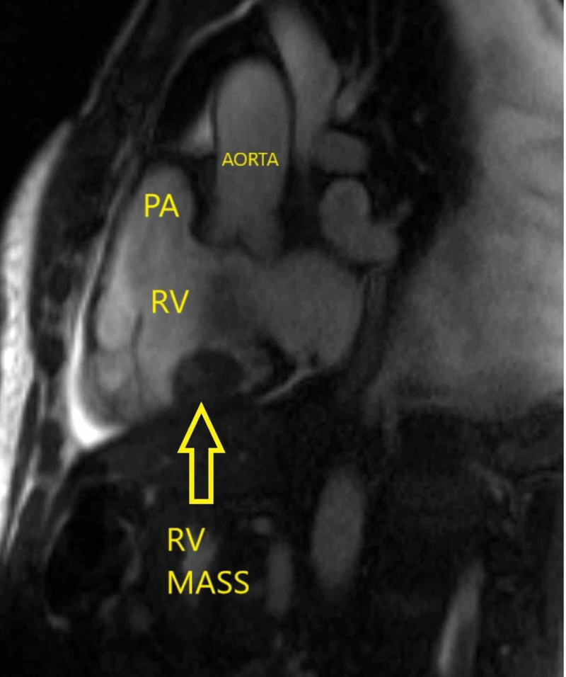
The presentation of fevers in a patient with active intravenous (IV) drug use is often challenging, as there is a wide range of both infectious and noninfectious disorders that can cause fevers. A thorough diagnostic workup is essential in identifying the etiology of these fevers. We report a rare case of an infected right ventricular (RV) thrombus as a cause of persistent fever and sepsis in a 46-year-old patient with IV drug use. The patient continued to have persistent bacteremia inspite of appropriate IV antibiotics. Hence, the patient warranted a cardiothoracic surgical excision of the infected RV thrombus following which the patient showed remarkable improvement.
Keywords: cocaine; infected cardiac thrombus; intravenous drug user; mssa bacteremia; sepsis; surgical resection.
Copyright © 2020, Arumairaj et al.
Conflict of interest statement
The authors have declared that no competing interests exist.
Figures
References
-
- Echocardiographic detection of an infected superior vena caval thrombus presenting as a right atrial mass. Dick AE, Gross CM, Rubin JW. Chest. 1989;96:212–214. - PubMed
-
- Infection of left ventricular thrombus in a patient with silent myocardial infarction—a unique complication. Senior R, Raftery EB. Eur Heart J. 1993;14:997–998. - PubMed
Publication types
LinkOut - more resources
Full Text Sources