. 2021 Jan;141(1):25-37.
doi: 10.1007/s00401-020-02233-8. Epub 2020 Nov 5.

Lewy pathology of the esophagus correlates with the progression of Lewy body disease: a Japanese cohort study of autopsy cases

Affiliations

Lewy pathology of the esophagus correlates with the progression of Lewy body disease: a Japanese cohort study of autopsy cases

Zen-Ichi Tanei et al. Acta Neuropathol. 2021 Jan.

Abstract

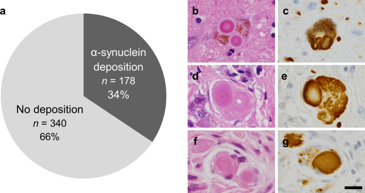
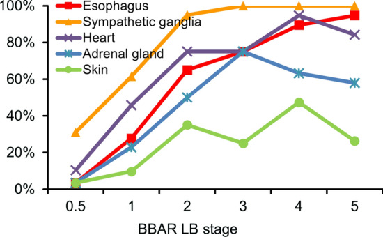
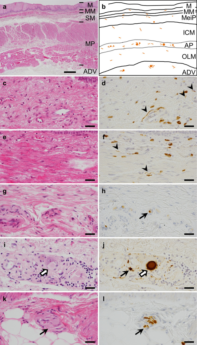
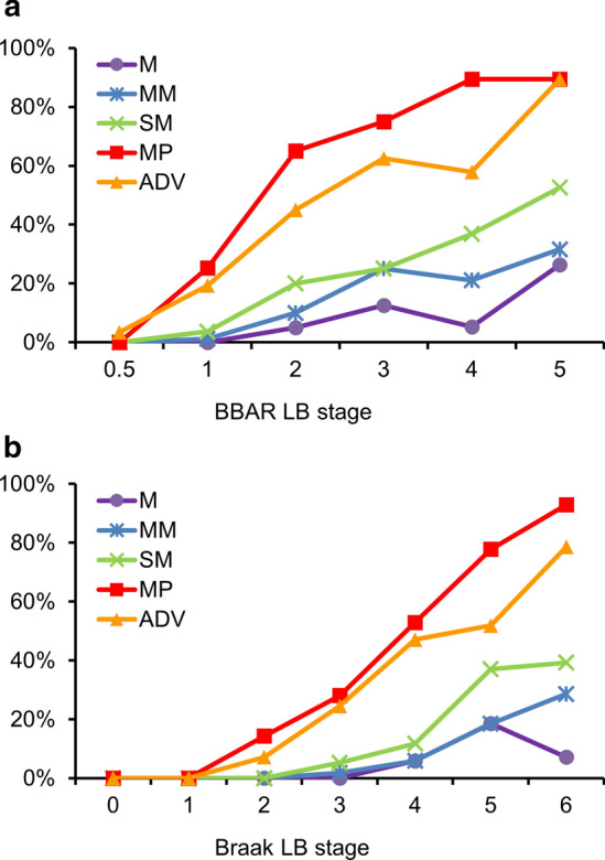
Lewy body disease (LBD) is a spectrum of progressive neurodegenerative disorders characterized by the wide distribution of Lewy bodies and neurites in the central and peripheral nervous system (CNS, PNS). Clinical diagnoses include Parkinson's disease (PD), dementia with Lewy bodies, or pure autonomic failure. All types of LBD are accompanied by non-motor symptoms (NMSs) including gastrointestinal dysfunctions such as constipation. Its relationship to Lewy body-related α-synucleinopathy (Lewy pathology) of the enteric nervous system (ENS) is attracting attention because it can precede the motor symptoms. To clarify the role of ENS Lewy pathology in disease progression, we performed a clinicopathological study using the Brain Bank for Aging Research in Japan. Five-hundred and eighteen cases were enrolled in the study. Lewy pathology of the CNS and PNS, including the lower esophagus as a representative of the ENS, was examined via autopsy findings. Results showed that one-third of older people (178 cases, 34%) exhibited Lewy pathology, of which 78 cases (43.8%) exhibited the pathology in the esophagus. In the esophageal wall, Auerbach's plexus (41.6%) was most susceptible to the pathology, followed by the adventitia (33.1%) and Meissner's plexus (14.6%). Lewy pathology of the esophagus was significantly associated with autonomic failures such as constipation (p < 0.0001) and among PNS regions, correlated the most with LBD progression (r = 0.95, p < 0.05). These findings suggest that the propagation of esophageal Lewy pathology is a predictive factor of LBD.

Keywords: Enteric nervous system; Esophagus; Lewy body disease; Parkinson’s disease; Peripheral nervous system; α-Synuclein.


Conflict of interest statement

The authors declare that they had no conflict of interest.

Figures

Fig. 1
Fig. 1
Prevalence of Lewy pathology in the 518 BBAR cases. a One-third of older individuals exhibited α-synuclein accumulation. bg LB in the locus coeruleus (b, c), adrenal gland (d, e), and esophagus (f, g). H&E (b, d, f) and pSyn#64 (c, e, g). Scale bar = 10 μm. BBAR the Brain Bank for Aging Research
Fig. 2
Fig. 2
Lewy pathology of the PNS at different BBAR LB stages. The positive rate of Lewy PNS pathology increased with the BBAR LB stage. Among PNS regions, the correlation with the BBAR LB stage was highest in the esophagus (r = 0.95), followed by the sympathetic ganglia (r = 0.85), heart (r = 0.87), adrenal gland (r = 0.81), and skin (r = 0.71; Spearman’s rank correlation coefficient, all p < 0.05). Positivity in the esophagus gradually increased with stage, while that in the heart and adrenal gland decreased at the last stage. The sympathetic ganglia exhibited the highest rate of pathology among the regions in the PNS. Details are summarized in Supplementary Table 2, online resource. BBAR the Brain Bank for Aging Research, LB Lewy body, PNS peripheral nervous system
Fig. 3
Fig. 3
Lewy pathology in the esophageal wall. a Low magnification view of the esophageal wall and its anatomy. b Schema of Lewy pathology, shown in orange spherical or neuritic structures in the esophageal wall. cl High magnification view of the mucosa, especially lamina propria (c, d), muscularis mucosa (e, f), submucosa (g, h), muscularis propria (i, j) and adventitia (k, l). LB were observed in Auerbach’s plexus in the MP (white arrows, i, j) and ADV. Lewy neurites or pSyn#64-immunoreactive (IR) aggregates were found in Meissner’s plexus of the SM, Auerbach’s plexus, and the ADV (arrows, h, j, k, l). Only pSyn#64-IR neurites were observed in the M and MM (arrowheads, d, f). Non-specific staining of stromal cell cytoplasm—most were mast cell granules—was observed in the background (d, f). H&E (a, c, e, g, i, k) and pSyn#64 (d, f, h, j, l). Scale bar = 500 µm (a), 20 µm (cl). LB Lewy body, M mucosa, MM muscularis mucosa, SM submucosa, MeiP Meissner’s plexus, MP muscularis propria, ICM inner circular muscle layer of MP, OLM outer longitudinal muscle layer of MP, AP Auerbach’s plexus, ADV adventitia
Fig. 4
Fig. 4
Lewy pathology in the esophageal wall at different BBAR and Braak LB stages. a BBAR LB stages; The positive rate of Lewy pathology increased with BBAR LB stage. The highest rates were found in the MP and ADV (mean rates of 41.6% and 33.1% respectively), reaching 89.5% at stage 5. Lewy pathology was observed less in the M and MM. b Braak LB stages; The positivity of Lewy pathology also increased with Braak LB stage. The highest rates were found in the MP, reaching 92.9% at stage 6. Lewy pathology was observed less in the M and MM. Details are summarized in Table 4a, b. BBAR the Brain Bank for Aging Research, LB Lewy body, M mucosa, MM muscularis mucosa, SM submucosa, MP muscularis propria, ADV adventitia

References

    1. Abbott RD, Petrovitch H, White LR, Masaki KH, Tanner CM, Curb JD, et al. Frequency of bowel movements and the future risk of Parkinson’s disease. Neurology. 2001;57:456–462. doi: 10.1212/wnl.57.3.456. - DOI - PubMed
    1. Annerino DM, Arshad S, Taylor GM, Adler CH, Beach TG, Greene JG. Parkinson’s disease is not associated with gastrointestinal myenteric ganglion neuron loss. Acta Neuropathol. 2012;124:665–680. doi: 10.1007/s00401-012-1040-2. - DOI - PMC - PubMed
    1. Baba M, Nakajo S, Tu PH, Tomita T, Nakaya K, Lee VM, et al. Aggregation of alpha-synuclein in Lewy bodies of sporadic Parkinson’s disease and dementia with Lewy bodies. Am J Pathol. 1998;152:879–884. - PMC - PubMed
    1. Beach TG, Adler CH, Sue LI, Vedders L, Lue L, White Iii CL, et al. Multi-organ distribution of phosphorylated alpha-synuclein histopathology in subjects with Lewy body disorders. Acta Neuropathol. 2010;119:689–702. doi: 10.1007/s00401-010-0664-3. - DOI - PMC - PubMed
    1. Beach TG, Corbille AG, Letournel F, Kordower JH, Kremer T, Munoz DG, et al. Multicenter assessment of immunohistochemical methods for pathological alpha-synuclein in sigmoid colon of autopsied Parkinson’s disease and control subjects. J Parkinsons Dis. 2016;6:761–770. doi: 10.3233/jpd-160888. - DOI - PMC - PubMed

Publication types

MeSH terms

Substances