Diagnostic value of optic disc retinal nerve fiber layer thickness for diabetic peripheral neuropathy
- PMID: 33150774
- PMCID: PMC7691684
- DOI: 10.1631/jzus.B2000225
Diagnostic value of optic disc retinal nerve fiber layer thickness for diabetic peripheral neuropathy
Abstract
Objective: To investigate the value of optic disc retinal nerve fiber layer (RNFL) thickness in the diagnosis of diabetic peripheral neuropathy (DPN).
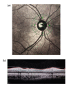
Methods: Ninety patients with type 2 diabetes, including 60 patients without DPN (NDPN group) and 30 patients with DPN (DPN group), and 30 healthy participants (normal group) were enrolled. Optical coherence tomography (OCT) was used to measure the four quadrants and the overall average RNFL thickness of the optic disc. The receiver operator characteristic curve was drawn and the area under the curve (AUC) was calculated to evaluate the diagnostic value of RNFL thickness in the optic disc area for DPN.
Results: The RNFL thickness of the DPN group was thinner than those of the normal and NDPN groups in the overall average ((101.07± 12.40) µm vs. (111.07±6.99) µm and (109.25±6.90) µm), superior quadrant ((123.00±19.04) µm vs. (138.93±14.16) µm and (134.47±14.34) µm), and inferior quadrant ((129.37±17.50) µm vs. (143.60±12.22) µm and (144.48±14.10) µm), and the differences were statistically significant. The diagnostic efficiencies of the overall average, superior quadrant, and inferior quadrant RNFL thicknesses, and a combined index of superior and inferior quadrant RNFL thicknesses were similar, and the AUCs were 0.739 (95% confidence interval (CI) 0.635-0.826), 0.683 (95% CI 0.576-0.778), 0.755 (95% CI 0.652-0.840), and 0.773 (95% CI 0.672-0.854), respectively. The diagnostic sensitivity of RNFL thickness in the superior quadrant reached 93.33%.
Conclusions: The thickness of the RNFL in the optic disc can be used as a diagnostic method for DPN.
Keywords: Type 2 diabetes; Peripheral neuropathy; Retinal nerve fiber layer thickness; Optical coherence tomography; Diagnosis.
Conflict of interest statement
All procedures followed were in accordance with the ethical standards of the responsible committee on human experimentation (institutional and national) and with the Helsinki Declaration of 1975, as revised in 2008 (5). This study was approved by the Ethics Committee of the Second Affiliated Hospital of Fujian Medical University, Quanzhou, China. Informed consent was obtained from all patients for being included in the study.
Figures


Similar articles
-
Optical coherence tomography of the retina combined with color Doppler ultrasound of the tibial nerve in the diagnosis of diabetic peripheral neuropathy.Front Endocrinol (Lausanne). 2022 Oct 21;13:938659. doi: 10.3389/fendo.2022.938659. eCollection 2022. Front Endocrinol (Lausanne). 2022. PMID: 36339439 Free PMC article.
-
Diagnostic capability of retinal thickness measures in diabetic peripheral neuropathy.J Optom. 2017 Oct-Dec;10(4):215-225. doi: 10.1016/j.optom.2016.05.003. Epub 2016 Jul 14. J Optom. 2017. PMID: 27423690 Free PMC article.
-
[Significance of optic disc topography and retinal nerve fiber layer thickness measurement by spectral-domain OCT in diagnosis of glaucoma].Zhonghua Yan Ke Za Zhi. 2010 Aug;46(8):702-8. Zhonghua Yan Ke Za Zhi. 2010. PMID: 21054994 Chinese.
-
Presence of Peripheral Neuropathy Is Associated With Progressive Thinning of Retinal Nerve Fiber Layer in Type 1 Diabetes.Invest Ophthalmol Vis Sci. 2017 May 1;58(6):BIO234-BIO239. doi: 10.1167/iovs.17-21801. Invest Ophthalmol Vis Sci. 2017. PMID: 28828484
-
Glaucoma diagnostic ability of quadrant and clock-hour neuroretinal rim assessment using cirrus HD optical coherence tomography.Invest Ophthalmol Vis Sci. 2012 Apr 24;53(4):2226-34. doi: 10.1167/iovs.11-8689. Invest Ophthalmol Vis Sci. 2012. PMID: 22410556
Cited by
-
Therapeutic effect of opioid analgesics combined with non-steroidal anti-inflammatory drugs on peripheral neuropathy and its influence on inflammatory factors.Am J Transl Res. 2021 Oct 15;13(10):11752-11757. eCollection 2021. Am J Transl Res. 2021. PMID: 34786103 Free PMC article.
-
Optical coherence tomography of the retina combined with color Doppler ultrasound of the tibial nerve in the diagnosis of diabetic peripheral neuropathy.Front Endocrinol (Lausanne). 2022 Oct 21;13:938659. doi: 10.3389/fendo.2022.938659. eCollection 2022. Front Endocrinol (Lausanne). 2022. PMID: 36339439 Free PMC article.
-
An Investigation of the Correlation Between Retinal Nerve Fiber Layer Thickness with Blood Biochemical Indices and Cognitive Dysfunction in Patients with Type 2 Diabetes Mellitus.Diabetes Metab Syndr Obes. 2024 Sep 4;17:3315-3323. doi: 10.2147/DMSO.S470297. eCollection 2024. Diabetes Metab Syndr Obes. 2024. PMID: 39247429 Free PMC article.
References
MeSH terms
Substances
LinkOut - more resources
Full Text Sources
Medical

