. 2021 Feb;30(2):350-365.
doi: 10.1002/pro.3990. Epub 2020 Nov 23.

Frontotemporal dementia-linked P112H mutation of TDP-43 induces protein structural change and impairs its RNA binding function

Affiliations

Frontotemporal dementia-linked P112H mutation of TDP-43 induces protein structural change and impairs its RNA binding function

Sashank Agrawal et al. Protein Sci. 2021 Feb.

Abstract

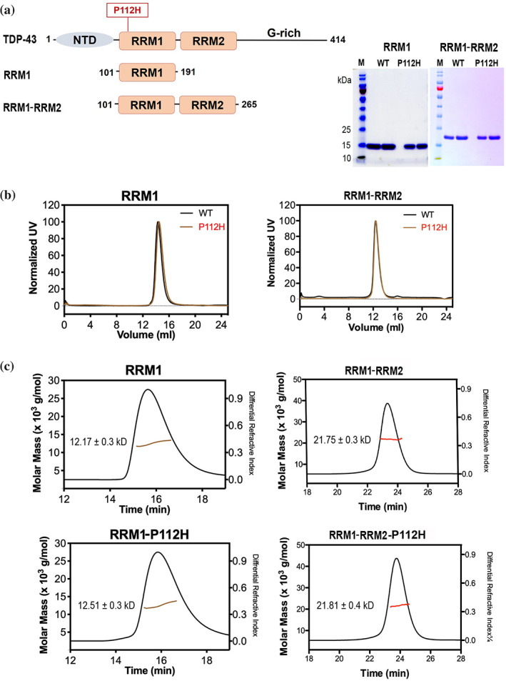
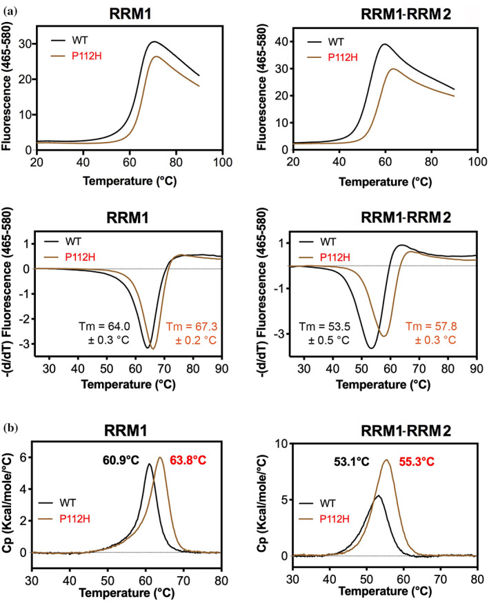
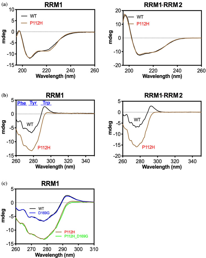
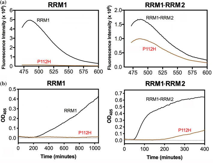
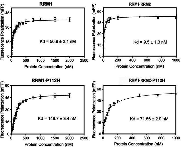
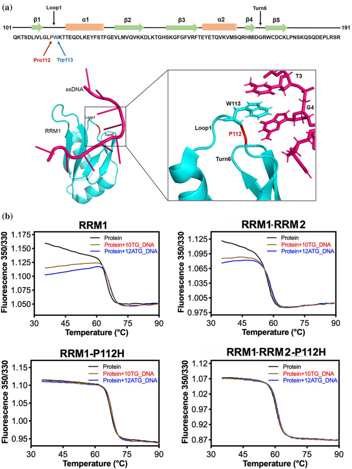
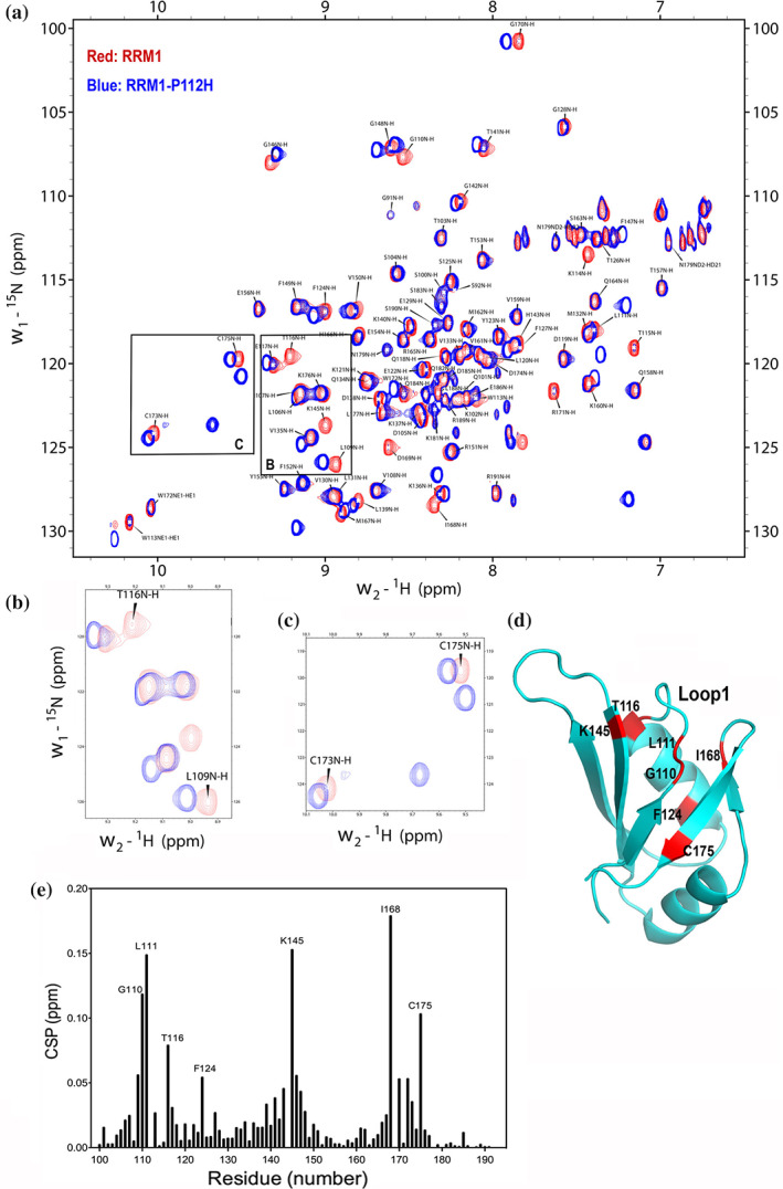
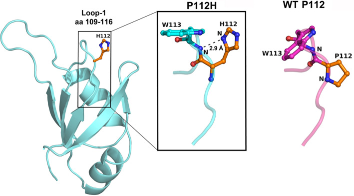
TDP-43 forms the primary constituents of the cytoplasmic inclusions contributing to various neurodegenerative diseases, including amyotrophic lateral sclerosis and frontotemporal dementia (FTD). Over 60 TDP-43 mutations have been identified in patients suffering from these two diseases, but most variations are located in the protein's disordered C-terminal glycine-rich region. P112H mutation of TDP-43 has been uniquely linked to FTD, and is located in the first RNA recognition motif (RRM1). This mutation is thought to be pathogenic, but its impact on TDP-43 at the protein level remains unclear. Here, we compare the biochemical and biophysical properties of TDP-43 truncated proteins with or without P112H mutation. We show that P112H-mutated TDP-43 proteins exhibit higher thermal stability, impaired RNA-binding activity, and a reduced tendency to aggregate relative to wild-type proteins. Near-UV CD, 2D-nuclear-magnetic resonance, and intrinsic fluorescence spectrometry further reveal that the P112H mutation in RRM1 generates local conformational changes surrounding the mutational site that disrupt the stacking interactions of the W113 side chain with nucleic acids. Together, these results support the notion that P112H mutation of TDP-43 contributes to FTD through functional impairment of RNA metabolism and/or structural changes that curtail protein clearance.

Keywords: RNA recognition motif; RNA-binding protein; neurodegenerative disease; protein aggregation.


Figures

FIGURE 1
FIGURE 1
TDP‐43 P112H mutants exhibit similar oligomeric states to wild‐type proteins. (a) The domain organization of full‐length TDP‐43, and the two truncated proteins, RRM1 and RRM1‐RRM2. The SDS‐PAGE (right panel) reveals that recombinant RRM1 and RRM1‐RRM2 proteins with or without P112H mutation had a high homogeneity. (b) Gel filtration (Superdex 75 10/300 GL column) profiles for the His‐tagged recombinant RRM1 and RRM2 with (red) or without P112H (black) mutation. (c) The molar mass of TDP‐43 proteins, estimated by size exclusion chromatography‐coupled multi‐angle light scattering (SEC‐MALS): RRM1 (12.17 kD), RRM1‐P112H (12.51 kD), RRM1‐RRM2 (21.75 kD), and RRM1‐RRM2‐P112H (21.81 kD). The calculated molecular weights for RRM1 and RRM1‐RRM2 are 12.4 and 20.8 kD, respectively
FIGURE 2
FIGURE 2
Mutation of P112H in TDP‐43 increases protein thermal stability. (a) The thermal melting points of wild‐type (in black) and P112H‐mutated (in red) TDP‐43 were assayed by differential scanning fluorimetry using SYPRO Orange dye as the fluorophore. The fluorescence signal, excited at 465 nm and emitted at 580 nm, was recorded while increasing the temperature from 20 to 90°C (upper panels). The melting point of each protein was estimated based on the derivatives of the fluorescence signal against temperature (shown in the lower panels and reflecting three independent measurements). (b) The thermal melting points of wild‐type TDP‐43 (in black) and P112H mutants (in red) were estimated by differential scanning calorimetry
FIGURE 3
FIGURE 3
Circular dichroism (CD) reveals conformational changes induced by P112H mutation in TDP‐43. (a) The far‐UV CD spectra of wild‐type TDP‐43 proteins (in black) and P112H mutants (in red) were recorded from 190 to 260 nm at 25°C at a protein concentration of 10 μM in a buffer containing 10 mM phosphate (pH 7.5) in a quartz cell with 1‐mm path length. (b) The near‐UV CD signal (from 260 to 350 nm) for each protein was recorded at a high protein concentration of 2 mg/ml in PBS buffer at 25°C in a quartz cuvette with 10‐mm path length. The peak region (260–310 nm) arise from aromatic amino acids are marked in the near‐UV CD of RRM1. (c) The near‐UV CD spectra for RRM1, RRM1‐P112H, RRM1‐D169G, and RRM1‐P112H‐D169G. Significant changes were observed in the spectra of RRM1‐P112H (in red) and RRM1‐P112H‐D169G (green), but not in RRM1‐D169G (blue), relative to that of wild‐type RRM1 (black)
FIGURE 4
FIGURE 4
TDP‐43 P112H mutants are resistant to forming amyloid inclusions and oxidation‐induced aggregation. (a) Time‐course experiments showing that TDP‐43 RRM1 and RRM1‐RRM2 formed amyloid inclusions in Thioflavin‐T (ThT) binding assays upon agitation in a buffer containing 25 mM Tris–HCl (pH 8.0 for RRM1 or pH 9.0 for RRM1‐RRM2) and 100 mM NaCl. In contrast, RRM1‐P112H and RRM1‐RRM2‐P112H were resistant to formation of ThT‐positive inclusions under the same conditions. (b) TDP‐43 RRM1 and RRM1‐RRM2 formed oxidation‐induced aggregates upon treatment with 5 mM H2O2, as revealed by the turbidity assay, whereas RRM1‐P112H and RRM1‐RRM2‐P112H formed significantly less oxidation‐induced aggregates under the same conditions
FIGURE 5
FIGURE 5
P112H mutation of TDP‐43 reduces RNA‐binding activity. The RNA‐binding affinities of TDP‐43 RRM1 and RRM1‐RRM2 proteins with or without P112H mutation for a 5′‐end FAM‐labeled (UG)6 RNA were measured by fluorescence polarization assays. The dissociation constants (Kd) and standard deviations shown in the figure were estimated from three independent measurements
FIGURE 6
FIGURE 6
P112H mutation of TDP‐43 RRM1 disrupts the interactions between Trp113 and nucleic acids. (a) The P112H mutation is located in Loop1 of RRM1. The crystal structure of TDP‐43 RRM1‐DNA complex (PDB entry: 4IUF) shows that the side chain of Trp113 is sandwiched between two nucleobases, making π‐π stacking interactions with DNA. 43 (b) Intrinsic tryptophan fluorescence profiles (ratio of absorption at 350/330 nm) of wild‐type RRM1 and RRM1‐RRM2 reveal diminished signal upon binding of a 10‐nt (10TG_DNA) or 12‐nt (12ATG_DNA) DNA (upper panels). However, RRM1‐P112H and RRM1‐RRM2‐P112H presented similar intrinsic tryptophan fluorescence profiles (lower panels) in the presence or absence of DNA, indicating that the environment surrounding residue Trp113 was not changed upon DNA binding
FIGURE 7
FIGURE 7
2D‐NMR reveals that the P112H mutation perturbs TDP‐43 RRM1 conformation. (a) Superimposition of the 15N‐HSQC NMR spectra of wild‐type RRM1 (in red) and RRM1‐P112H (in blue), revealing that the P112H mutation induces peak shifts. The peak assignments for the wild‐type RRM1 spectrum were imported from BMRB entry 18,765. (b) A representative region shows that residues Leu109 and Thr116 located within Loop1 of RRM1‐P112H exhibit significant shifts of HSQC peaks. (c) A closer look at the HSQC spectrum highlights the peak shifts in two cysteine residues of RRM1‐P112H. (d) Amino acids exhibiting significant shifts of HSQC peaks (marked in red) occur mainly in two regions, Loop1 and Turn6, and are mapped onto the RRM1 structure. (e) The chemical shift perturbation (CSP) between the 15N‐HSQC spectra of RRM1 and RRM1‐P112H were calculated and plotted as a function of residue number. No value is shown for residues that could not be assigned in one or both spectra
FIGURE 8
FIGURE 8
Molecular model of TDP‐43 RRM1‐P112H mutant. The molecular model of RRM1‐P112H was built using the crystal structure of wild‐type RRM1 (PDB entry: 4Y0F) as the template. 43 The geometry of the initial RRM1‐P112H model was optimized using the modeling tool in PHENIX (version 1.13–2,298). In the final model, the side chain (Nδ1 atom) of His112 located in Loop1 forms a hydrogen bond with the main‐chain nitrogen of Trp113, suggesting that P112H mutation likely decreases the loop flexibility, and thus increases the thermal stability of TDP‐43

Similar articles

Cited by

References

    1. Vanden Broeck L, Callaerts P, Dermaut B. TDP‐43‐mediated neurodegeneration: Towards a loss‐of‐function hypothesis? Trends Mol Med. 2014;20:66–71. - PubMed
    1. Sephton CF, Cenik B, Cenik BK, Herz J, Yu G. TDP‐43 in central nervous system development and function: Clues to TDP‐43‐associated neurodegeneration. Biol Chem. 2012;393:589–594. - PMC - PubMed
    1. Butti Z, Patten SA. RNA dysregulation in amyotrophic lateral sclerosis. Front Genet. 2018;9:712. - PMC - PubMed
    1. Zhang YJ, Caulfield T, Xu YF, et al. The dual functions of the extreme N‐terminus of TDP‐43 in regulating its biological activity and inclusion formation. Hum Mol Genet. 2013;22:3112–3122. - PMC - PubMed
    1. Shiina Y, Arima K, Tabunoki H, Satoh J. TDP‐43 dimerizes in human cells in culture. Cell Mol Neurobiol. 2010;30:641–652. - PMC - PubMed

Publication types