. 2020 Nov 25;83(11):3287-3297.
doi: 10.1021/acs.jnatprod.0c00570. Epub 2020 Nov 5.

Targeting Trimeric and Tetrameric Proanthocyanidins of Cinnamomum verum Bark as Bioactives for Dental Therapies

Affiliations

Targeting Trimeric and Tetrameric Proanthocyanidins of Cinnamomum verum Bark as Bioactives for Dental Therapies

Joo-Won Nam et al. J Nat Prod. .

Abstract

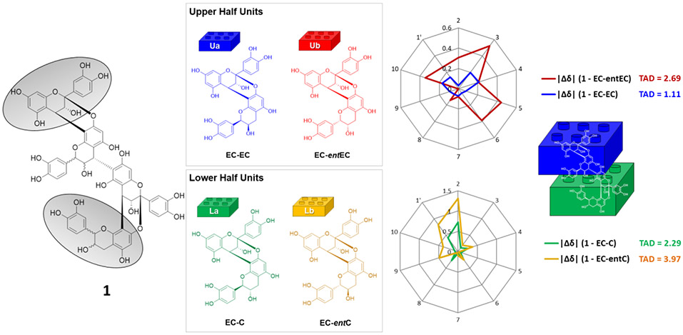
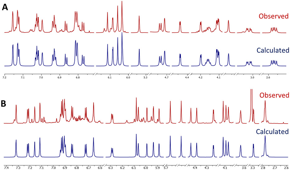
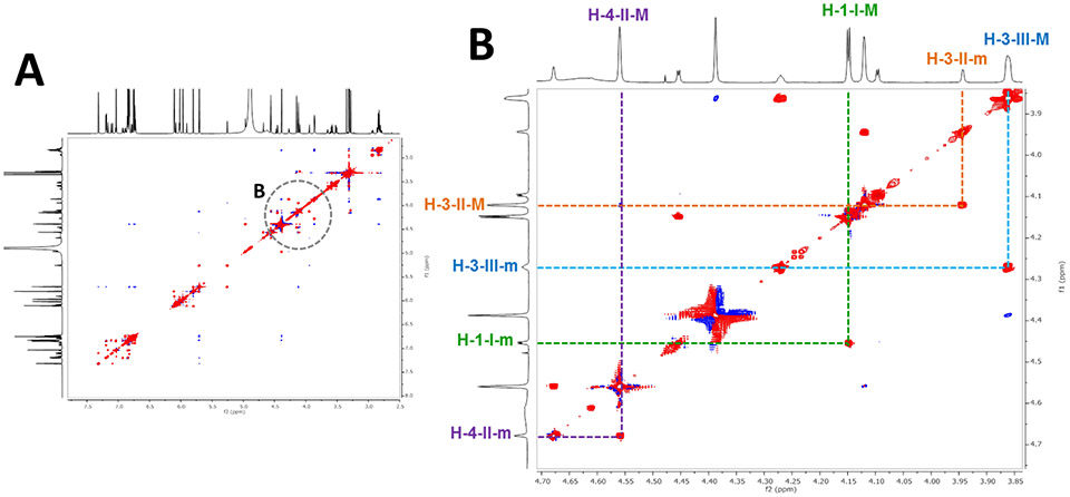
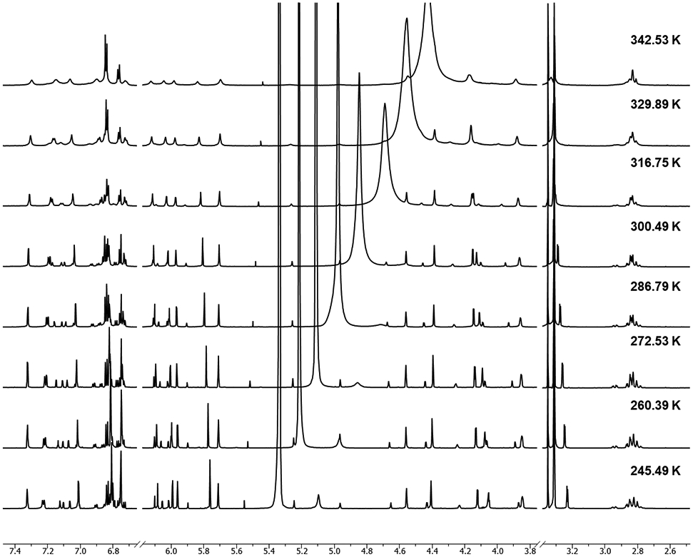
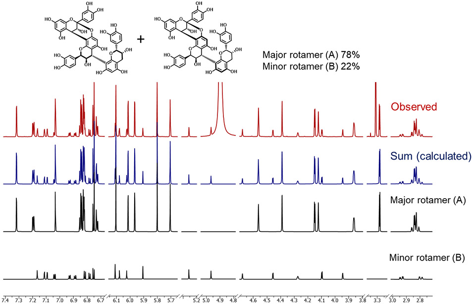
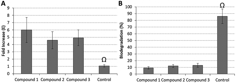
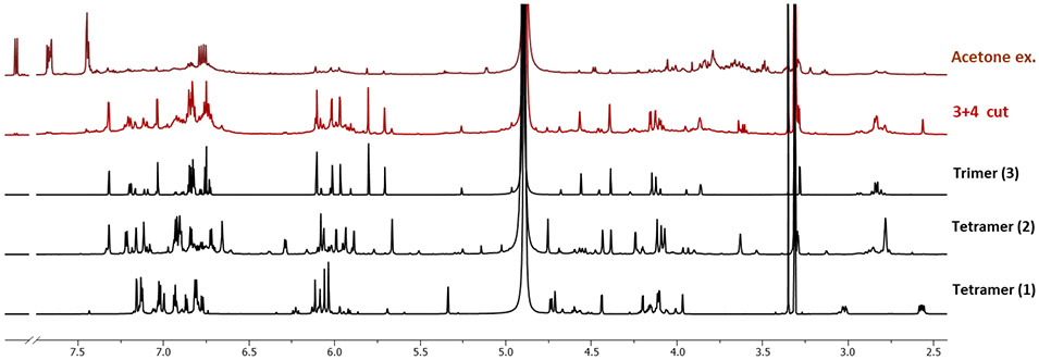
The present study elucidated the structures of three A-type tri- and tetrameric proanthocyanidins (PACs) isolated from Cinnamomum verum bark to the level of absolute configuration and determined their dental bioactivity using two therapeutically relevant bioassays. After selecting a PAC oligomer fraction via a biologically diverse bioassay-guided process, in tandem with centrifugal partition chromatography, phytochemical studies led to the isolation of PAC oligomers that represent the main bioactive principles of C. verum: two A-type tetrameric PACs, epicatechin-(2β→O→7,4β→8)-epicatechin-(4β→6)-epicatechin-(2β→O→7,4β→8)-catechin (1) and parameritannin A1 (2), together with a trimer, cinnamtannin B1 (3). Structure determination of the underivatized proanthocyanidins utilized a combination of HRESIMS, ECD, 1D/2D NMR, and 1H iterative full spin analysis data and led to NMR-based evidence for the deduction of absolute configuration in constituent catechin and epicatechin monomeric units.


Figures

Figure 1.
Figure 1.
Structures of the oligomeric proanthocyanidins (OPACs), 1-3, isolated from C. verum bark, in comparison with their core A-type dimer, proanthocyanidin A2. In addition to the classical chemical formulas, the graphical PAC Block ARray representations (PACBAR; bottom) along with the text-based macro and micro PACBAR codes.
Figure 2.
Figure 2.
Determination of the absolute configuration of 1 via comparison of 13C chemical shift differences with those of its four stereochemically possible A-type dimeric PAC moieties.
Figure 3.
Figure 3.
The congruence of the observed (red, methanol-d4, 900 MHz) and calculated (blue) 1H NMR spectra of OPACs 1 (A) and 2 (B) demonstrates the validity of the HiFSA profiles of the two isolates.
Figure 4.
Figure 4.
The ROESY spectrum (900 MHz) shows exchange correlation cross peaks between the two atropisomers of 3 giving rise to signals that exhibit the same phase as the diagonal (red; “in-phase” cross peaks). The major and minor atropisomers are denoted as M and m, respectively.
Figure 5.
Figure 5.
Stacked 1H NMR spectra of 3 acquired at various temperatures in methanol-d4 (800 MHz).
Figure 6.
Figure 6.
Quantitative and qualitative 1H NMR HiFSA-based determination of the major (A) and minor rotamers (B) of 3. The experimental (observed, red) spectrum was acquired at 900 MHz in methanol-d4.
Figure 7.
Figure 7.
Biomodification potential by two relevant bioassays: (A) fold increase observed in the apparent modulus of elasticity (E) of the dentin matrix; (B) biodegradation rates (in %) of dentin specimens treated with 13. The Ω symbol depicts statistically significant differences between groups treated with 1-3 and control for both E (p ≤ 0.001) and biodegradation (p < 0.001).
Figure 8.
Figure 8.
The 1H NMR spectra of a 70% acetone extract, a trimer and tetramer enriched fraction (3+4 cut), and the isolates 1-3 from C. verum. The fraction 3+4 cut (red) was prepared in a prior study and its 1H NMR spectrum is shown for comparison. The 1H NMR spectra of the 70% acetone extract and the 3+4 cut were acquired under the same conditions (concentration, volume, NMR acquisition parameters, 600 MHz, methanol-d4). The 1H NMR spectra of 1-3 were acquired at 900 MHz in methanol-d4.

References

    1. Vidal CMP; Leme AA; Aguiar TR; Phansalkar RS; Nam J-W; Bisson J; McAlpine JB; Chen S-N; Pauli GF; Bedran-Russo AK, Langmuir 2014, 30, 14887–14893. - PMC - PubMed
    1. He L; Mu C; Shi J; Zhang Q; Shi B; Lin W, Int. J. Biol. Macromol 2011, 48, 354–359. - PubMed
    1. Bedran-Russo AK; Pashley DH; Agee K; Drummond JL; Miescke KJ, J. Biomed. Mater. Res. Part B Appl. Biomater 2008, 86, 330–334. - PubMed
    1. Aydin B; Leme-Kraus AA; Vidal CMP; Aguiar TR; Phansalkar RS; Nam J-W; McAlpine JB; Chen S-N; Pauli GF; Bedran-Russo AK, Dent. Mater 2019, 35, 328–334. - PMC - PubMed
    1. Aguiar TR; Vidal CMP; Phansalkar RS; Todorova I; Napolitano JG; McAlpine JB; Chen SN; Pauli GF; Bedran-Russo AK, J. Dent. Res 2014, 93, 417–422. - PMC - PubMed

Publication types