. 2020 Nov 5;15(11):e0241007.
doi: 10.1371/journal.pone.0241007. eCollection 2020.

MiR-21 mediates the protection of kaempferol against hypoxia/reoxygenation-induced cardiomyocyte injury via promoting Notch1/PTEN/AKT signaling pathway

Affiliations

MiR-21 mediates the protection of kaempferol against hypoxia/reoxygenation-induced cardiomyocyte injury via promoting Notch1/PTEN/AKT signaling pathway

Jinxi Huang et al. PLoS One. .

Abstract

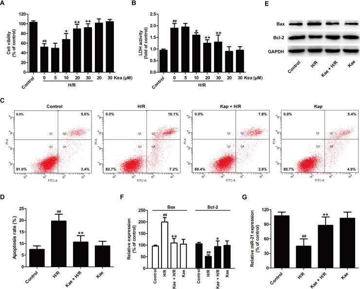
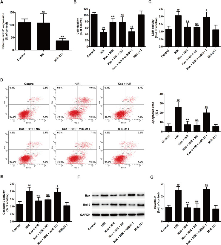
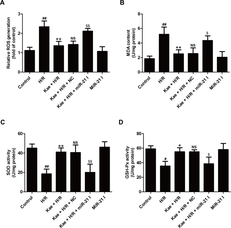
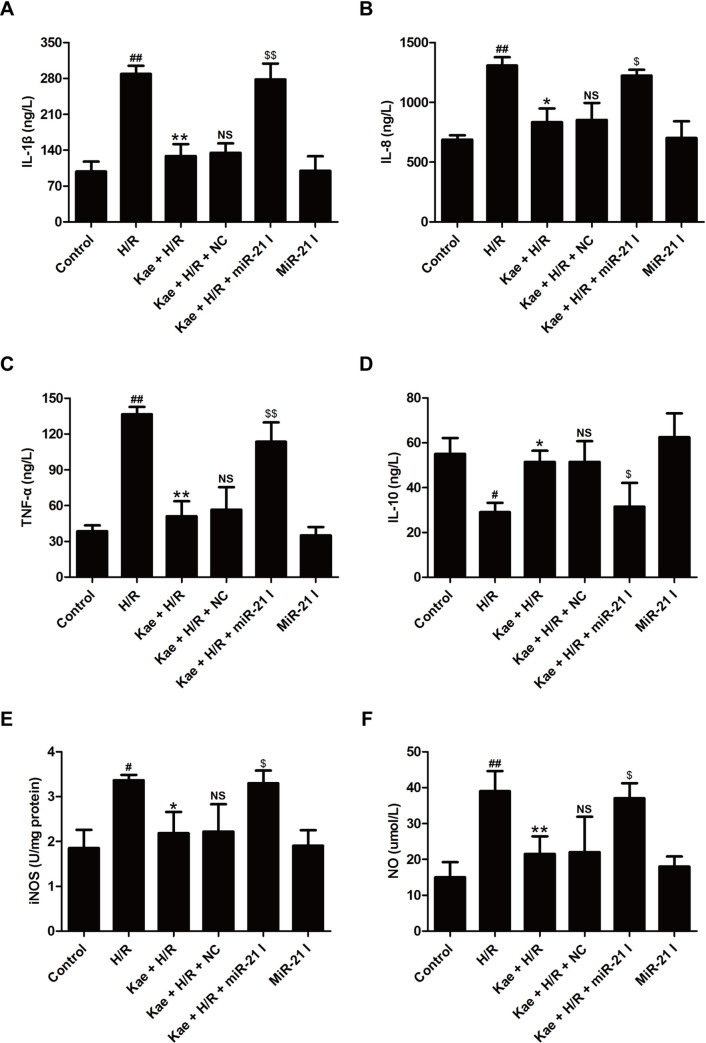
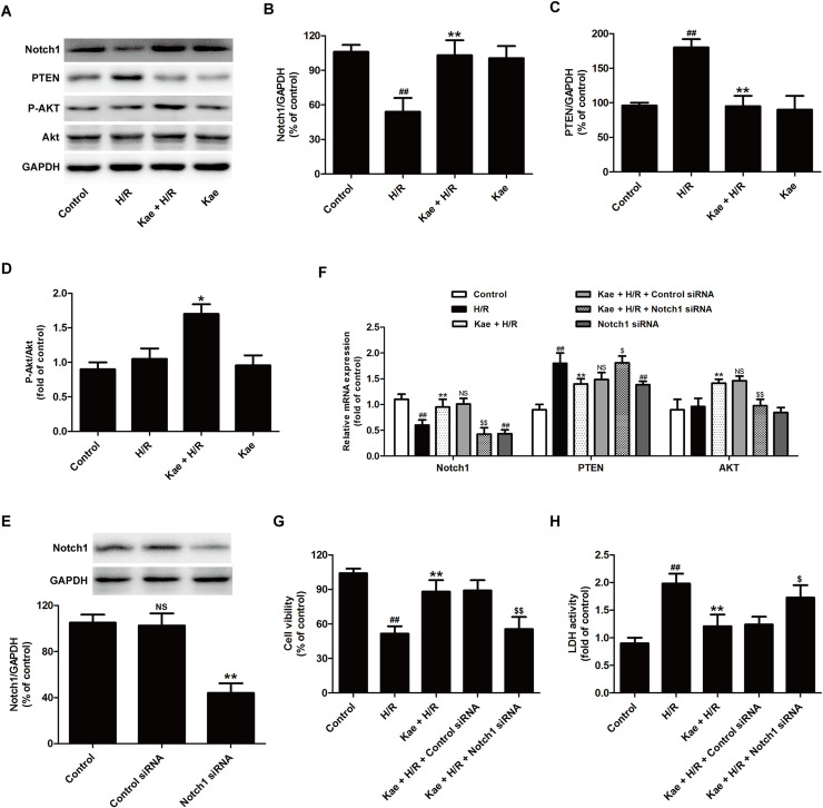
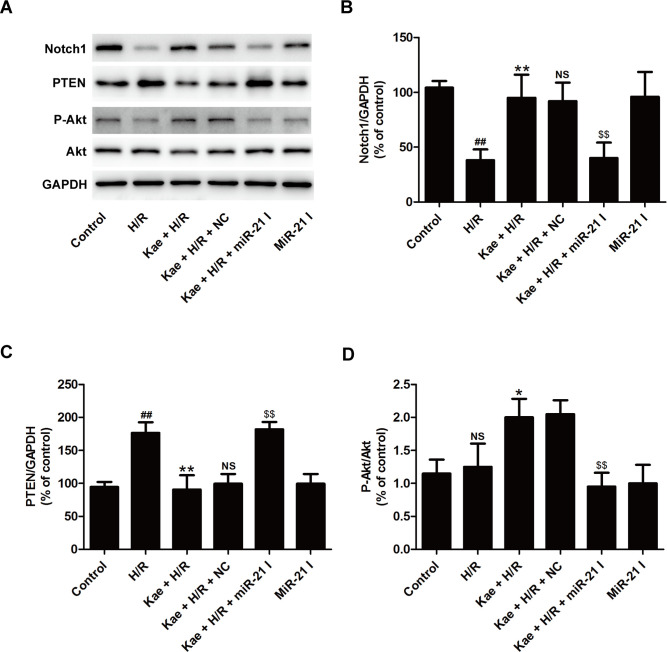
Kaempferol, a natural flavonoid compound, possesses potent myocardial protective property in ischemia/reperfusion (I/R), but the underlying mechanism is not well understood. The present study was aimed to explore whether miR-21 contributes to the cardioprotective effect of kaempferol on hypoxia/reoxygenation (H/R)-induced H9c2 cell injury via regulating Notch/phosphatase and tensin homologue (PTEN)/Akt signaling pathway. Results revealed that kaempferol obviously attenuates H/R-induced the damages of H9c2 cells as evidence by the up-regulation of cell viability, the down-regulation of lactate dehydrogenase (LDH) activity, the reduction of apoptosis rate and pro-apoptotic protein (Bax) expression, and the increases of anti-apoptotic protein (Bcl-2) expression. In addition, kaempferol enhanced miR-21 level in H9c2 cells exposed to H/R, and inhibition of miR-21 induced by transfection with miR-21 inhibitor significantly blocked the protection of kaempferol against H/R-induced H9c2 cell injury. Furthermore, kaempferol eliminated H/R-induced oxidative stress and inflammatory response as illustrated by the decreases in reactive oxygen species generation and malondialdehyde content, the increases in antioxidant enzyme superoxide dismutase and glutathione peroxidase activities, the decreases in pro-inflammatory cytokines interleukin (IL)-1β, IL-8 and tumor necrosis factor-alpha levels, and an increase in anti-inflammatory cytokine IL-10 level, while these effects of kaempferol were all reversed by miR-21 inhibitor. Moreover, results elicited that kaempferol remarkably blocks H/R-induced the down-regulation of Notch1 expression, the up-regulation of PTEN expression, and the reduction of P-Akt/Akt, indicating that kaempferol promotes Notch1/PTEN/AKT signaling pathway, and knockdown of Notch1/PTEN/AKT signaling pathway induced by Notch1 siRNA also abolished the protection of kaempferol against H/R-induced the damage of H9c2 cells. Notably, miR-21 inhibitor alleviated the promotion of kaempferol on Notch/PTEN/Akt signaling pathways in H9c2 cells exposed to H/R. Taken together, these above findings suggested thatmiR-21 mediates the protection of kaempferol against H/R-induced H9c2 cell injuryvia promoting Notch/PTEN/Akt signaling pathway.


Conflict of interest statement

The authors have declared that no competing interests exist.

Figures

Fig 1
Fig 1. Kaempferol protects cardiomyocyte injury and increases miR-21 expression in H9c2 cell-subjected with H/R.
H9c2 cells were pretreated with kaempferol (5, 10, 20, and 30 μM) for 2 h and then co-treated with H (6 h)/R (12 h). (A) CCK-8 assay for cell viability. (B) LDH detection kit for LDH activity in cellular supernatant. Data are presented as the means ± SD, n = 3. Compared with the control group, ##P < 0.001; compared with the H/R group, *P < 0.05, **P < 0.01. H9c2 cells were pretreated with kae (20 μM) for 2 h and then co-treated with H (6 h)/R (12 h). (C) Annexin V-FITC staining assay for apoptosis rates. (D) A statistical representation of apoptosis rates. (E) Western blot analysis for Bax and Bcl-2 expression. (F) Quantitative analysis of Bax and Bcl-2 expressions normal to GAPDH. (G) RT-PCR for miR-21 level. Data are presented as the means ± SD, n = 3. Compared with the control group, #P < 0.05, ##P < 0.001; compared with the H/R group, *P < 0.05, **P < 0.01. Kae, kaempferol; H/R, hypoxia/reoxygenation.
Fig 2
Fig 2. MiR-21 inhibitor attenuates kaempferol-induced the inhibitory effects on H/R-induced cytotoxicity and apoptosis in H9c2 cells.
H9c2 cells were transfected with miR-21 inhibitor (miR-21 I) or negative control (NC). (A) RT-PCR for miR-21 level. Data are presented as the means ± SD, n = 3. Compared with the control group, NSP > 0.05; compared with the NC transfection group, **P < 0.01. H9c2 cells were transfected with miR-21 I or NC followed by treatment with kaempferol (20 μM) for 2 h and then co-treatment with H (6 h)/R (12 h). (B) CCK-8 assay for cell viability. (C) LDH detection kit for LDH activity in cellular supernatant. (D) Annexin V-FITC staining assay for apoptosis. (E) Determination of caspase-3 activity by commercial kit. (F) Western blot analysis for Bax and Bcl-2 expression. (G) Quantitative analysis of Bax/Bcl-2 normal to GAPDH. Data are presented as the means ± SD, n = 3. Compared with the control group, #P < 0.05, ##P < 0.001; compared with the H/R group, *P < 0.05, **P < 0.01; compared with the Kae + H/R group, NSP > 0.05; compared with the Kae + H/R + NC group, $P < 0.05, $$P < 0.01. NS: No significance. Kae, kaempferol; H/R, hypoxia/reoxygenation.
Fig 3
Fig 3. MiR-21 inhibitor reverses kaempferol-induced inhibition on oxidative stress in H9c2 cells exposed to H/R.
H9c2 cells were transfected with miR-21 inhibitor (miR-21 I) or negative control (NC) followed by treatment with kaempferol (20 μM) for 2 h and then co-treatment with H (6 h)/R (12 h). (A) ROS production was detected by 2’,7’-dichlorofluorescein diacetate (DCFH-DA). (B) Malondialdehyde (MDA) content, (C) superoxide dismutase (SOD), and (D) glutathione peroxidase (GSH-Px) were measured by corresponding commercial kits. Data are presented as the means ± SD, n = 3. Compared with the control group, #P < 0.05, ##P < 0.001; compared with the H/R group, *P < 0.05, **P < 0.01; compared with the Kae + H/R group, NSP > 0.05; compared with the Kae + H/R + NC group, $P < 0.05, $$P < 0.01.Kae, kaempferol; H/R, hypoxia/reoxygenation.
Fig 4
Fig 4. MiR-21 inhibitor blocks kaempferol-induced the inhibition on inflammatory response in H9c2 cells exposed to H/R.
H9c2 cells were transfected with miR-21 inhibitor (miR-21 I) or negative control (NC) followed by treatment with kaempferol (20 μM) for 2 h and then co-treatment with H (6 h)/R (12 h). The levels of IL-1β (A), IL-8 (B), TNF-α (C) and IL-10 (D), and the activity of iNOS were determined by ELISA, respectively. (F) The content of NO was analyzed by the Nitrate/Nitrite Assay Kit. Data are presented as the means ± SD, n = 3. Compared with the control group, #P < 0.05, ##P < 0.001; compared with the H/R group, *P < 0.05, **P < 0.01; compared with the Kae + H/R group, NSP > 0.05; compared with the Kae + H/R + NC group, $P < 0.05, $$P < 0.01. Kae, kaempferol; H/R, hypoxia/reoxygenation.
Fig 5
Fig 5. Kaempferol blocks H/R-induced the inhibition on Notch1/PTEN/Akt signaling pathway in H9c2 cells exposed to H/R.
H9c2 cells were pretreated with kaempferol (20 μM) for 2 h and then co-treated with H/R. (A) Protein expressions were measured by Western blot analysis. Quantitative analysis of Notch1 (B), PTEN (C), and P-Akt/Akt (D) normal to GAPDH. Data are presented as the means ± SD, n = 3. Compared with the control group, ##P < 0.01; compared with the H/R group, *P < 0.05, **P < 0.01. H9c2 cells were transfected with control siRNA and Notch1 siRNA, respectively. (E) Western blot analysis for Notch1 normal to GAPDH. Data are presented as the means ± SD, n = 3. Compared with the control group, NSP > 0.05; compared with the control siRNA group, **P < 0.01. H9c2 cells were transfected with control siRNA or Notch1 siRNA followed by treatment with kaempferol (20 μM) for 2 h and then co-treated with H/R. (F) RT-PCR for mRNA level and Quantitative analysis. (G) CCK-8 assay for cell viability. (H) LDH detection kit for LDH activity in cellular supernatant. Data are presented as the means ± SD, n = 3. Compared with the control group, ##P < 0.001; compared with the H/R group, **P < 0.01; compared with the Kae + H/R + control siRNA group, $P < 0.05, $$P < 0.01. Notch1, neurogenic locus notch homolog protein‑1; PTEN, phosphatase and tensin homolog deleted on chromosome ten.
Fig 6
Fig 6. MiR-21 inhibitor abolishes kaempferol-induced the activation of Notch1/PTEN/Akt signaling pathway in H9c2 cells exposed to H/R.
H9c2 cells were transfected with miR-21 inhibitor (miR-21 I)or negative control (NC) followed by treatment with kaempferol (20 μM) for 2 h and then co-treatment with H (6 h)/R (12 h). (A) Western blot analysis for protein expression. (B) Quantitative analysis of Notch1 expression (C), PTEN expression, and (D) P-Akt/Akt. Data are presented as the means ± SD, n = 3. Compared with the control group, #P < 0.05, ##P < 0.001; compared with the H/R group, *P < 0.05, **P < 0.01; compared with the Kae + H/R group, NSP > 0.05; compared with the Kae + H/R + NC group, $P < 0.05, $$P < 0.01.

Similar articles

Cited by

References

    1. Sanada S, Komuro I, Kitakaze M (2011) Pathophysiology of myocardial reperfusion injury: preconditioning, postconditioning, and translational aspects of protective measures. Am J Physiol Heart Circ Physiol 301: H1723–1741. 10.1152/ajpheart.00553.2011 - DOI - PubMed
    1. Buja LM (2005) Myocardial ischemia and reperfusion injury. Cardiovasc Pathol 14: 170–175. 10.1016/j.carpath.2005.03.006 - DOI - PubMed
    1. Ding J, Yang Z, Ma H, Zhang H (2019) Mitochondrial Aldehyde Dehydrogenase in Myocardial Ischemic and Ischemia-Reperfusion Injury. Adv Exp Med Biol 1193: 107–120. 10.1007/978-981-13-6260-6_6 - DOI - PubMed
    1. Sinning C, Westermann D, Clemmensen P (2017) Oxidative stress in ischemia and reperfusion: current concepts, novel ideas and future perspectives. Biomark Med 11: 11031–11040. 10.2217/bmm-2017-0110 - DOI - PubMed
    1. Thind GS, Agrawal PR, Hirsh B, Saravolatz L, Chen-Scarabelli C, et al. (2015) Mechanisms of myocardial ischemia-reperfusion injury and the cytoprotective role of minocycline: scope and limitations. Future Cardiol 11: 61–76. 10.2217/fca.14.76 - DOI - PubMed

MeSH terms