. 2020 Dec 2;48(21):12297-12309.
doi: 10.1093/nar/gkaa998.

PpCas9 from Pasteurella pneumotropica - a compact Type II-C Cas9 ortholog active in human cells

Affiliations

PpCas9 from Pasteurella pneumotropica - a compact Type II-C Cas9 ortholog active in human cells

Iana Fedorova et al. Nucleic Acids Res. .

Abstract

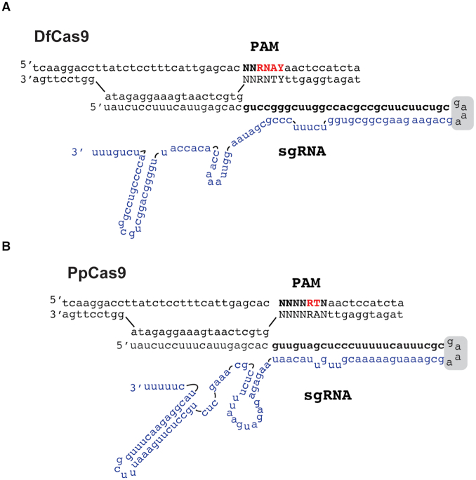
CRISPR-Cas defense systems opened up the field of genome editing due to the ease with which effector Cas nucleases can be programmed with guide RNAs to access desirable genomic sites. Type II-A SpCas9 from Streptococcus pyogenes was the first Cas9 nuclease used for genome editing and it remains the most popular enzyme of its class. Nevertheless, SpCas9 has some drawbacks including a relatively large size and restriction to targets flanked by an 'NGG' PAM sequence. The more compact Type II-C Cas9 orthologs can help to overcome the size limitation of SpCas9. Yet, only a few Type II-C nucleases were fully characterized to date. Here, we characterized two Cas9 II-C orthologs, DfCas9 from Defluviimonas sp.20V17 and PpCas9 from Pasteurella pneumotropica. Both DfCas9 and PpCas9 cleave DNA in vitro and have novel PAM requirements. Unlike DfCas9, the PpCas9 nuclease is active in human cells. This small nuclease requires an 'NNNNRTT' PAM orthogonal to that of SpCas9 and thus potentially can broaden the range of Cas9 applications in biomedicine and biotechnology.


Figures

Figure 1.
Figure 1.
Defluviimonas sp. 20V17 and Pasteurella pneumotropica CRISPR-Cas Type II-C loci. (A) Organization of Defluviimonas sp. 20V17 and P. pneumotropica CRISPR-Cas Type II-C loci. DRs are shown as black rectangles, spacers are indicated by rectangles of different colors. The tracrRNA coding sequences are shown as yellow rectangles. The cas genes are labeled. Direction of transcription is indicated with black arrows. (B) In silico co-folding of Defluviimonas sp. 20V17 and P. pneumotropica DRs and putative tracrRNAs. The DR sequences are colored in red, the tracrRNA sequences are colored in green.
Figure 2.
Figure 2.
Studying of Defluviimonas sp. 20V17 CRISPR-Cas Type II-C system in bacteria. (A) Identification of Defluviimonas sp. 20V17 crRNAs. Reads (blue) are mapped at the top of the CRISPR array. A sequence of a typical mature crRNA sequence is expanded at the bottom with spacer part shown in orange. The direction of transcription is indicated with black arrows. (B) Determination of DfCas9 PAM sequences using plasmid transformation interference screening. Above: a scheme of the interference screen experiment. Below: DfCas9 PAM sequence logo determined from the PAM screening. PAM position numbers correspond to nucleotides immediately following the protospacer in the 5′-3′ direction.
Figure 3.
Figure 3.
In vitro cleavage of DNA targets by DfCas9. (A) Single-nucleotide substitutions in the third, fourth and sixth positions of PAM prevent DNA cleavage by DfCas9. An agarose gel showing the results of electrophoretic separation of cleavage products of targets with PAM sequences shown at the top is presented. Bands corresponding to cleaved and uncleaved DNA fragments are indicated. The scheme above shows the position of expected DNA cleavage site. (B) DfCas9 cleaves different DNA targets with 5′-NNRNAYN-3′ PAM consensus in vitro. The scheme above shows the positions of different target sites in the grin2b gene fragment. Below: A gel showing results of in vitro cleavage of targets with indicated PAMs is presented.
Figure 4.
Figure 4.
In vitro cleavage of DNA targets by PpCas9. (A) A scheme of the in vitro PAM screening experiment. A linear DNA PAM library containing a target site flanked with seven randomized nucleotides at the 3′ end is incubated with PpCas9 charged with appropriate crRNA and tracrRNA. This leads to the cleavage of library members carrying functional PAM sequences and generates DNA products shortened by 50 bp. Uncleaved PAM library molecules are recovered, which depletes the library. The uncleaved molecules, as well as negative control (original PAM library incubated with PpCas9-tracrRNA in the absence of crRNA) are sequenced. Comparison of PAM variants representation in the depleted sample and the control allows to determine the PpCas9 PAM logo. (B) Web logo of PpCas9 PAM sequences depleted after in vitro PAM screening. (C) Wheel representation of in vitro PAM screen results for fifth, sixth and seventh nucleotide positions of PAM. Nucleotide positions from the inner to the outer circle match PAM positions moving away from the protospacer. For a given sequence, the area of the sector in the PAM wheel displays the relative depletion in the library. (D) Single-nucleotide substitutions in the fifth and sixth positions of PAM prevent DNA cleavage by PpCas9. An agarose gel showing the results of electrophoretic separation of targets with PAM sequences shown at the top after incubation during 30 min with the PpCas9 effector complex is presented. Bands corresponding to cleaved and uncleaved DNA fragments are indicated. (E) PpCas9 efficiently cleaves different DNA targets with 5′-NNNNRTN-3′ PAM consensus in vitro. The scheme above shows the positions of different target sites in a 1592 bp GRIN2b gene fragment. Below: A gel showing results of in vitro cleavage of targets with indicated PAMs is presented.
Figure 5.
Figure 5.
DNA cleavage activity of DfCas9 and PpCas9 at different temperatures. DfCas9 or PpCas9 was incubated with cognate tracrRNA and crRNA and a 2.7 kb plasmid DNA or a 921 bp linear DNA fragment containing target sequences at indicated temperatures for 10 min. Products were separated by agarose gel electrophoresis. Cleavage efficiency (in percent) was calculated as a ratio of intensity of cleaved bands to the combined intensity of cleaved and uncleaved bands. Mean values and standard deviations obtained from three independent experiments are shown.
Figure 6.
Figure 6.
The DfCas9 and PpCas9 minimal in vitro DNA cleavage systems. (A) A scheme of recognition by the DfCas9–sgRNA complex of a DNA target flanked by 5′-NNRNAY-3′ PAM (Y stands for pyrimidines, R stands for purines). The crRNA-tracrRNA linker is indicated with a gray box. The part of sgRNA that originated from tracrRNA is shown in blue. (B) A scheme of recognition by the PpCas9–sgRNA complex of a DNA target flanked by 5′-NNNNRTN-3 PAM (R stands for purines). The crRNA-tracrRNA linker is indicated by a gray box. The part of sgRNA that originated from tracrRNA is shown in blue.
Figure 7.
Figure 7.
PpCas9 nuclease activity in human HEK293T cells. (A) Scheme of the PpCas9 nuclease activity assessment experiment. Above: A scheme showing design of a plasmid used for PpCas9 gene and sgRNAs expression. The PpCas9 gene is shown as a yellow rectangle, NLS (nuclear localization signals) as brown rectangles, GFP gene as a green rectangle. CMV promoter and U6 promoters are indicated with black arrows. The sgRNA coding sequence is shown as a green rectangle. The plasmid was transfected into HEK293T cells and genomic DNA was extracted from a heterogeneous population of modified and unmodified cells for indel frequency assessment through HTS of the targeted region or in vitro assay with T7 endonuclease I. (B) Results of T7 endonuclease I indel detection assay showing PpCas9-mediated cleavage of EMX1 and GRIN2b genes in HEK293T genome. (C) PpCas9 indel formation efficiency at different genomic sites. Left: genomic DNA target sites with corresponding PAM sequences. 5′-NNNNRTT-3′ PAM are shown in red. Right: indel frequency estimated by HTS analysis. Mean values and standard deviations obtained from three biological replicas are shown. (D) The influence of sgRNA spacer length on PpCas9-mediated indel formation efficiency in EMX1 and GRIN2b genes. HEK293T cells were transfected with PpCas9_sgRNA plasmids (as in panel A) coding for sgRNAs with different lengths of spacer segments. Left: results for the GRIN2b.1 target, right: for the EMX1.1 target. Mean values and standard deviations obtained from three biological replicas are shown.
Figure 8.
Figure 8.
The specificity of genomic DNA cleavage by PpCas9. The indel frequency at two on-target as well as at corresponding off-target sequences was assessed by targeted amplicon sequencing of genomic DNA of HEK293T cells transfected with plasmids carrying the PpCas9 genome editing system. Left: Sequences of on-target sites (in green) and off-target sites are shown. Mismatches in off-target sequences are shown in red. Right: Frequencies of indel formation in each site (mean values and standard deviations obtained from three replicas are shown in a table).
Figure 9.
Figure 9.
PpCas9 is closely related to NmeCas9 and PmCas9. (A) An unrooted phylogenetic tree showing the relationship of PpCas9 to NmeCas9 and PmCas9. Nodes with strong statistical support (bootstrap values > 80%) are shown. (B) An alignment of PpCas9, PmCas9 and NmeCas9 direct repeat sequences. Identical nucleotides are indicated with asterisks. (C) Comparison of CRISPR-Cas Type II-C loci from Pasteurella pneumotropica, Pasteurella multocida Pm70 and Neisseria meningitidis. The percent of amino acid identities with P. pneumotropica sequences for Cas1, Cas2 and Cas9 orthologues and nucleotide identities for DRs are shown. (D) PpCas9, PmCas9 and NmeCas9 tracrRNA coding sequences alignment. Identical nucleotides are indicated with asterisks.

Similar articles

Cited by

References

    1. Barrangou R., Fremaux C., Deveau H., Richards M., Boyaval P., Moineau S., Romero D.A., Horvath P.. CRISPR provides acquired resistance against viruses in prokaryotes. Science. 2007; 315:1709–1712. - PubMed
    1. Garneau J.E., Dupuis M.-È., Villion M., Romero D.A., Barrangou R., Boyaval P., Fremaux C., Horvath P., Magadán A.H., Moineau S.. The CRISPR/Cas bacterial immune system cleaves bacteriophage and plasmid DNA. Nature. 2010; 468:67–71. - PubMed
    1. Marraffini L.A., Sontheimer E.J.. CRISPR interference limits horizontal gene transfer in staphylococci by targeting DNA. Science. 2008; 322:1843–1845. - PMC - PubMed
    1. Jansen R., van Embden J.D.A., Gaastra W., Schouls L.M.. Identification of genes that are associated with DNA repeats in prokaryotes. Mol. Microbiol. 2002; 43:1565–1575. - PubMed
    1. Pourcel C., Salvignol G., Vergnaud G.. CRISPR elements in Yersinia pestis acquire new repeats by preferential uptake of bacteriophage DNA, and provide additional tools for evolutionary studies. Microbiology (Reading). 2005; 151:653–663. - PubMed

Publication types

MeSH terms

Substances