. 2021 Mar 1;148(5):1245-1259.
doi: 10.1002/ijc.33383. Epub 2020 Dec 2.

Targeting stromal cell Syndecan-2 reduces breast tumour growth, metastasis and limits immune evasion

Affiliations

Targeting stromal cell Syndecan-2 reduces breast tumour growth, metastasis and limits immune evasion

Paul G Loftus et al. Int J Cancer. .

Abstract

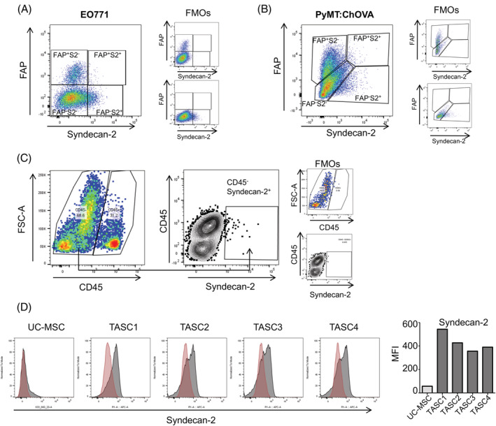
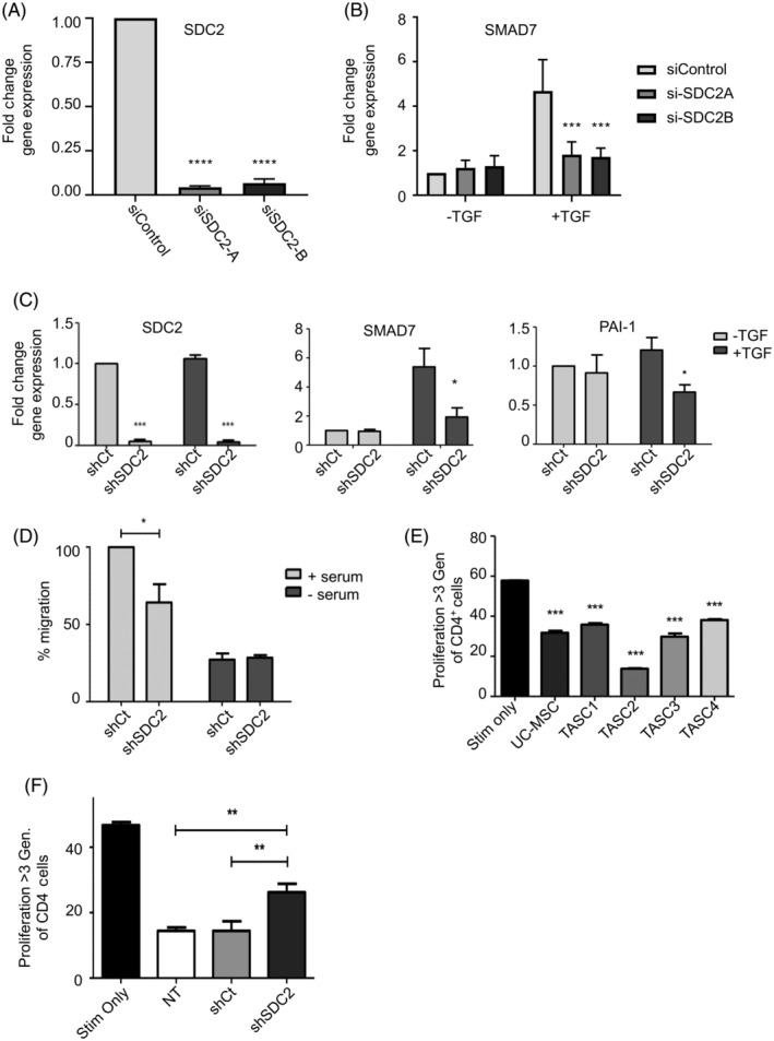
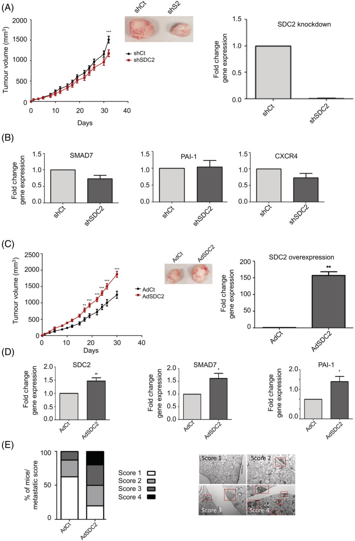
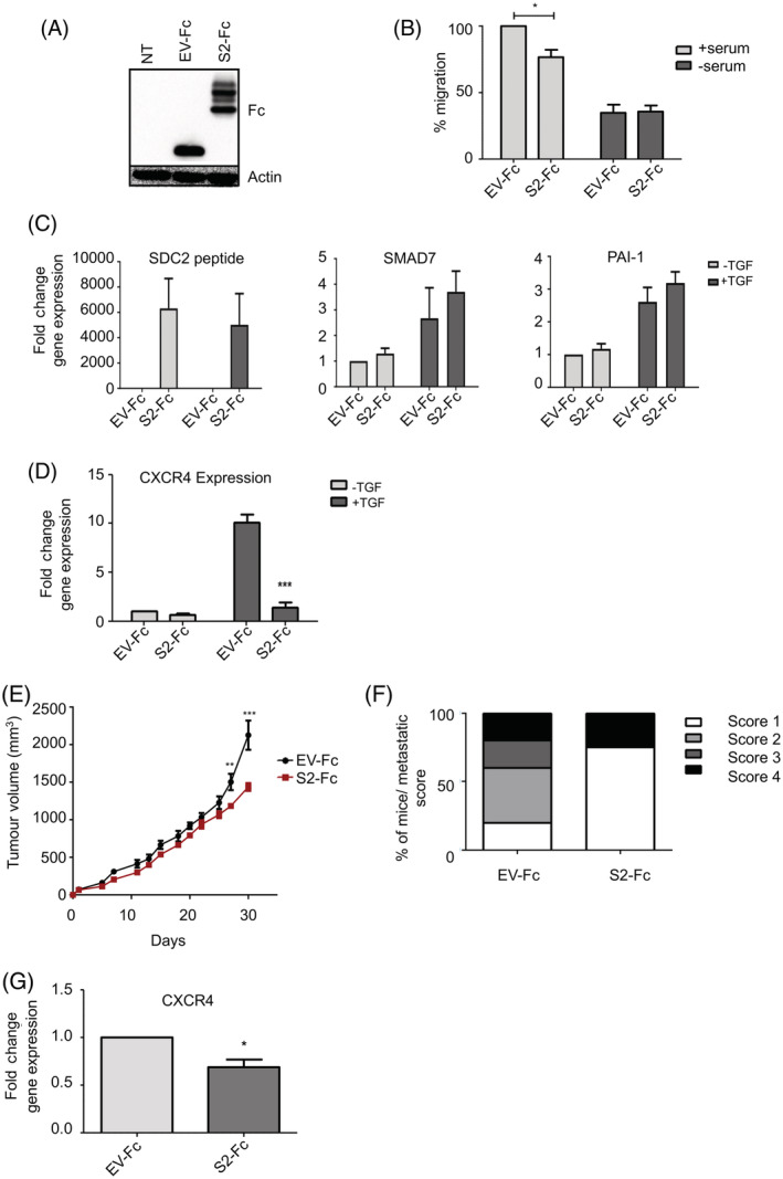
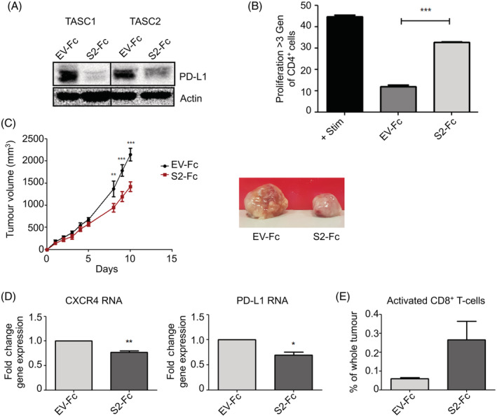
Tumour stromal cells support tumourigenesis. We report that Syndecan-2 (SDC2) is expressed on a nonepithelial, nonhaematopoietic, nonendothelial stromal cell population within breast cancer tissue. In vitro, syndecan-2 modulated TGFβ signalling (SMAD7, PAI-1), migration and immunosuppression of patient-derived tumour-associated stromal cells (TASCs). In an orthotopic immunocompromised breast cancer model, overexpression of syndecan-2 in TASCs significantly enhanced TGFβ signalling (SMAD7, PAI-1), tumour growth and metastasis, whereas reducing levels of SDC2 in TASCs attenuated TGFβ signalling (SMAD7, PAI-1, CXCR4), tumour growth and metastasis. To explore the potential for therapeutic application, a syndecan-2-peptide was generated that inhibited the migratory and immunosuppressive properties of TASCs in association with reduced expression of TGFβ-regulated immunosuppressive genes, such as CXCR4 and PD-L1. Moreover, using an orthotopic syngeneic breast cancer model, overexpression of syndecan-2-peptide in TASCs reduced tumour growth and immunosuppression within the TME. These data provide evidence that targeting stromal syndecan-2 within the TME inhibits tumour growth and metastasis due to decreased TGFβ signalling and increased immune control.

Keywords: Fc-peptide; TGFβ signalling; breast cancer; immunosuppression; syndecan-2; tumour-associated stromal cells.


Conflict of interest statement

TOB is founder, director and shareholder of Orbsen Therapeutics Ltd. SJE, PL, LW LMD and SA are employees and shareholders of Orbsen Therapeutics Ltd. LOF is a former employee and shareholder of Orbsen Therapeutics Ltd. MG, MK, RMD, ECR and LRB have no conflicts of interest to declare.

Figures

FIGURE 1
FIGURE 1
Syndecan‐2+ stromal cells within breast tumours. A, EO771 tumours were dissected and single cell suspensions stained with Syndecan‐2, FAP, CD45, Ter119, CD31 and EpCAM antibodies. TASCs were defined by flow cytometry as being CD45 (nonlymphocyte) Ter119 (nonerythroid), EpCAM (nonepithelial) and CD31 (nonendothelial) cells. The levels of viable (Sytox ‐ve) Syndecan‐2+ stromal (CD45Ter199CD31EpCAM) were determined. Fluorescence minus one (FMO) controls were used to identify and gate cells. B, 24‐week‐old PyMT:ChOVA spontaneous tumours were dissected and single cell suspensions stained as earlier. C, Human breast cancer samples were digested and single cell suspensions stained with Syndecan‐2 and CD45 antibodies to determine the levels of viable (Sytox ‐ve) Syndecan‐2+/CD45 and Syndecan‐2+/CD45+ cells by flow cytometry. D, Flow cytometry histograms and mean fluorescence intensity (MFI) illustrating cell surface expression of Syndecan‐2 [Color figure can be viewed at wileyonlinelibrary.com]
FIGURE 2
FIGURE 2
Modulation of SDC2 effects TGFβ signalling, migration and immunosuppressive properties of TASCs. A, RT‐qPCR analysis showing siRNA‐SDC2 transfected TASCs (siSDC2‐A and siSDC2‐B) have reduced SDC2 RNA levels compared to siControl transfected TASCs. B, siControl or siSDC2 transfected TASCs were treated with TGFβ. 2 hours after TGFβ treatment RNA was prepared and levels of SMAD7 were determined by RT‐qPCR. n = 3; ***P ≤ .0001. C, shControl (shCt) or shSDC2 transduced TASCs were treated with TGFβ. 2 hours after TGFβ treatment RNA was prepared and levels of SDC2 and TGFβ‐regulated genes were determined by RT‐qPCR. n = 4; *P ≤ .05 ***P ≤ .0001. D, The xCelligence system was used to determine the ability of shCt and shSDC2 transduced TASCs to migrate towards serum‐containing media. Levels of migration were normalised to the positive control (ie, shCt +serum). n = 4 *P ≤ .05. E, TASCs and umbilical cord MSCs (UC‐MSCs) were cocultured with CD3/CD28‐activated peripheral blood mononuclear cells (PBMCs) at a 1:50 ratio. Flow cytometry of CFSE‐labelled CD4+ T cells reveal CD3/CD28‐mediated proliferation is inhibited by TASCs. One‐way analysis of variance (ANOVA) with Tukey's multiple comparison test ***P ≤ .001. F, shCt or shSDC2 transduced TASCs were cocultured with CD3/CD28‐activated peripheral blood mononuclear cells (PBMCs) at a ratio of 1:50. Flow cytometry of CFSE‐labelled CD4+ T cells was used to measure the level of proliferation. Data were compared by one‐way ANOVA with Tukey's multiple comparison posttest. n = 4 (**P ≤ .01)
FIGURE 3
FIGURE 3
Manipulation of SDC2 within the stromal cell compartment of xenograft tumours effects breast carcinogenesis. A, Orthotopic xenograft tumours were established by coinjecting MDA‐MB‐231 with shSDC2‐transduced TASCs or shCt‐transduced TASCs into the mammary fat pad of immune‐compromised mice at a ratio of 1:10 (TASCs:MDA). Tumours containing shSDC2‐TASCs (n = 9) showed significantly lower growth rates when compared to control shCt‐TASC tumours (n = 10). RT‐qPCR analysis showing shSDC2‐transduced TASCs have reduced SDC2 RNA levels compared to adenovirus shCt‐transduced TASCs. B, RNA was prepared from xenograft tumours and RT‐qPCR was performed to compare the expression of TGFβ‐regulated genes between shCt and shSDC2 expressing tumours (n = 5/group). C, Orthotopic xenograft tumours were established as described earlier. Tumours containing TASCs overexpressing SDC2 (AdSDC2) (n = 10) showed significantly higher growth rates when compared to control tumours (AdCt) (n = 9). RT‐qPCR analysis showing AdSDC2‐transduced TASCs have increased SDC2 RNA levels compared to adenovirus AdCt‐transduced TASCs. D, RNA was prepared from xenograft tumours and RT‐qPCR was performed to determine the effect of SDC2 modulation within TASCs upon the expression of TGFβ‐regulated genes. (n = 5/group). E, Approximately 12 weeks after TASC:MDA injection, lungs were removed and examined for metastatic nodules by H&E staining. (AdCt [n = 9], AdSDC2 [n = 10]). The bar graph indicates the metastatic score/lung from the various experimental groups. Red boxes indicate metastatic lesions. *P ≤ .05; **P ≤ .01; ***P ≤ .001 [Color figure can be viewed at wileyonlinelibrary.com]
FIGURE 4
FIGURE 4
Syndecan‐2 peptide decreases breast tumour growth and TGFβ signalling. A, Western blot analysis demonstrating overexpression of Fc‐tagged syndecan‐2 peptide (S2‐Fc) and Fc‐empty vector control (EV‐Fc) in TASCs compared to nontransfected TASCs (NT). B, The xCelligence system was used to determine the ability of EV‐Fc‐ and S2‐Fc‐transfected TASCs to migrate towards serum‐containing media. Levels of migration were normalised to the positive control (ie, EV‐Fc + serum). C, EV‐Fc or S2‐Fc expressing TASCs were treated with TGFβ, RNA was prepared and levels of TGFβ‐regulated genes were determined by RT‐qPCR, n = 4. D, EV‐Fc or S2‐Fc expressing TASCs were treated with TGFβ, following treatment RNA was prepared and levels of CXCR4 were determined by RT‐qPCR. E, NOD:SCID tumours containing TASCs expressing S2‐Fc show a significant decrease in tumour growth compared EV‐Fc control tumours (n = 5 [EV‐Fc] n = 4 [S2‐Fc]). F, Lungs were removed and examined for metastatic nodules by H&E staining. The bar graph indicates the metastatic score/lung from the two experimental groups. G, Tumours were excised and RT‐qPCR was performed to determine the levels of CXCR4 expression. *P ≤ .05; **P ≤ .01; ***P ≤ .001 [Color figure can be viewed at wileyonlinelibrary.com]
FIGURE 5
FIGURE 5
Syndecan‐2 peptide reduces the immunosuppressive properties of TASCs, reduces tumour growth and increases the percentage of activated T cells within the TME. A, TASCs were transfected with EV‐Fc control or S2‐Fc, 48 hours later protein extracts were prepared and PD‐L1 levels were determined by Western blot analysis. B, EV‐Fc or S2‐Fc expressing TASCs were cocultured with CD3/CD28‐activated peripheral blood mononuclear cells (PBMCs) at a ratio of 1:10. Flow cytometry of CFSE‐labelled CD4+ T cells was used to measure the level of proliferation. Data were compared by one‐way analysis of variance with Tukey's multiple comparison posttest. n = 3. C, EO771 tumours containing TASCs expressing S2‐Fc show a significant decrease in tumour growth compared EV‐Fc control tumours (n = 4 [EV‐Fc] n = 4 [F2‐Fc]). D, Tumours were excised and RT‐qPCR was performed to determine the levels of CXCR4 and PD‐L1. E, Flow cytometry analysis of tumours shows trends towards an increase in the number of CD8+, CD4, CD62Llo, CD44hi and CD25+‐activated T cells. *P ≤ .05; **P ≤ .01; ***P ≤ .001 [Color figure can be viewed at wileyonlinelibrary.com]

References

    1. Siegel RL, Miller KD, Jemal A. Cancer statistics, 2018. CA Cancer J Clin. 2018;68:7‐30. - PubMed
    1. Arendt LM, Rudnick JA, Keller PJ, Kuperwasser C. Stroma in breast development and disease. Semin Cell Dev Biol. 2010;21:11‐18. - PMC - PubMed
    1. Tchou J, Conejo‐Garcia J. Targeting the tumor stroma as a novel treatment strategy for breast cancer: shifting from the neoplastic cell‐centric to a stroma‐centric paradigm. Adv Pharmacol. 2012;65:45‐61. - PubMed
    1. Farmer P, Bonnefoi H, Anderle P, et al. A stroma‐related gene signature predicts resistance to neoadjuvant chemotherapy in breast cancer. Nat Med. 2009;15:68‐74. - PubMed
    1. Roodhart JM, Daenen LG, Stigter EC, et al. Mesenchymal stem cells induce resistance to chemotherapy through the release of platinum‐induced fatty acids. Cancer Cell. 2011;20:370‐383. - PubMed

Publication types

MeSH terms