Review
. 2020 Nov 3;9(11):2402.
doi: 10.3390/cells9112402.

Glutamatergic Receptor Trafficking and Delivery: Role of the Exocyst Complex

Affiliations
Review

Glutamatergic Receptor Trafficking and Delivery: Role of the Exocyst Complex

Matías Lira et al. Cells. .

Abstract

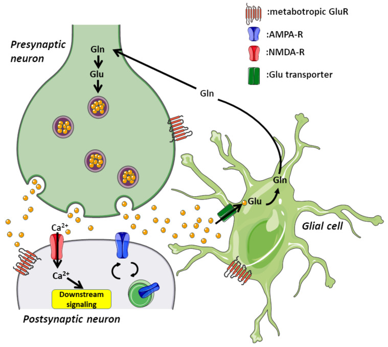
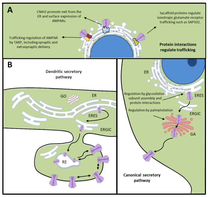
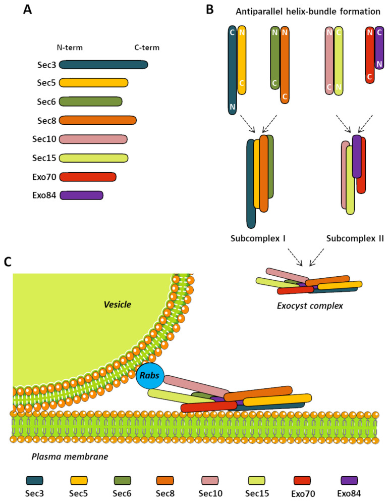
Cells comprise several intracellular membrane compartments that allow them to function properly. One of these functions is cargo movement, typically proteins and membranes within cells. These cargoes ride microtubules through vesicles from Golgi and recycling endosomes to the plasma membrane in order to be delivered and exocytosed. In neurons, synaptic functions employ this cargo trafficking to maintain inter-neuronal communication optimally. One of the complexes that oversee vesicle trafficking and tethering is the exocyst. The exocyst is a protein complex containing eight subunits first identified in yeast and then characterized in multicellular organisms. This complex is related to several cellular processes, including cellular growth, division, migration, and morphogenesis, among others. It has been associated with glutamatergic receptor trafficking and tethering into the synapse, providing the molecular machinery to deliver receptor-containing vesicles into the plasma membrane in a constitutive manner. In this review, we discuss the evidence so far published regarding receptor trafficking and the exocyst complex in both basal and stimulated levels, comparing constitutive trafficking and long-term potentiation-related trafficking.

Keywords: LTP-induced trafficking; constitutive trafficking; exocyst; glutamatergic receptor; membrane trafficking; synapse.


Conflict of interest statement

The authors declare no conflict of interest.

Figures

Figure 1
Figure 1
Excitatory synapse structure. Excitatory synapses are the most common in the central nervous system. It comprises a presynaptic neuron, a postsynaptic neuron, and a glial cell. The presynaptic compartment contains the cellular machinery for tethering, exocytosis, and endocytosis of glutamate-containing vesicles; refilling these vesicles is carried out in this compartment as well. In the postsynaptic compartment, we find glutamate receptors and their associated signaling coupled to exocytosis and recycling machinery. Additionally, there are supporting glial cells that maintain this basic synaptic structure. The correct functioning of synapses depends on all three cellular components in order to maintain physiological activity.
Figure 2
Figure 2
Ionotropic glutamate receptor trafficking. (A) Protein interactions regulate exocytic trafficking of ionotropic glutamate receptors, such as TARPs, Cornichon proteins, and scaffolding proteins, among others. (B) (Left) Dendritic secretory pathway, indicating the presence of ER, ERES, ERGIC, and GO in the dendrites. Moreover, it shows the action of recycling endosomes in the anterograde secretory pathway for glutamate receptors to the plasma membrane. (Right) Canonical secretory pathway for glutamatergic receptors in the somatic ER, and trafficking through ERGIC and GA. Neurons use both pathways in neuronal development and plasticity. ER: endoplasmic reticulum, ERES: endoplasmic reticulum exits sites, ERGIC: ER–Golgi intermediate compartment, GO: Golgi outpost, GA: Golgi apparatus, TARP: transmembrane α-amino-3-hydroxy-5-methyl-4-isoxazole propionic acid receptor regulatory proteins.
Figure 3
Figure 3
The exocyst complex. (A) Bars are examples of the scaled size corresponding to each subunit. (B) The scheme depicts antiparallel helix-bundle formation in the assembly of the exocyst complex. Subunit positioning corresponds to an approximation of the interaction experiments discussed in this review. Primary pairs are formed by Sec3–Sec5, Sec6–Sec8, Sec10–Sec15, and Exo70–Exo84. These pairs interact closely to induce the formation of the subcomplexes 1 and 2. Then, both subcomplexes bind together to finally stabilize the functional exocyst complex. (C) A cartoon of the exocyst complex is shown. The exocyst acts by tethering secretory vesicles to the plasma membrane in specific sites where the cargo is needed. The model shows a rod-like molecular structure of the subunits. In this model, Sec3 and Exo70 target the exocyst to the plasma membrane. An opposite positioning of Sec10 and Sec15 is observed compared to Sec3 and Exo70; this positioning is vital to the contact of the exocyst with the vesicles where Sec15 interacts with several Rabs to be its effector protein.
Figure 4
Figure 4
Exocyst in glutamatergic receptors trafficking. (Left) In basal conditions, AMPARs are trafficked along with the GRIP1 scaffold protein and the Sec8 subunit of the exocyst complex. NMDARs, on the other hand, are trafficked in vesicles along with SAP102 scaffold protein and subunits of the exocyst complex such as Sec8, Sec6, and Exo70. (Right) It is believed that the LTP-driven trafficking of glutamate receptors is augmented. In this context, the role of the exocyst complex is unknown. Nevertheless, the exocyst has been suggested as the only tethering complex at the plasma membrane in several cell types. Through its intrinsic exocytic faculties, it would participate in LTP-induced trafficking and the exocytosis of glutamate receptors. LTP: long-term potentiation.

References

    1. Meldrum B.S. Glutamate as a neurotransmitter in the brain: Review of physiology and pathology. J. Nutr. 2000;130:1007S–1015S. doi: 10.1093/jn/130.4.1007S. - DOI - PubMed
    1. Hering H., Sheng M. Dendritic spines: Structure, dynamics and regulation. Nat. Rev. Neurosci. 2001;2:880–888. doi: 10.1038/35104061. - DOI - PubMed
    1. Hackett J.T., Ueda T. Glutamate Release. Neurochem. Res. 2015;40:2443–2460. doi: 10.1007/s11064-015-1622-1. - DOI - PubMed
    1. Blackshaw L.A., Page A.J., Young R.L. Metabotropic glutamate receptors as novel therapeutic targets on visceral sensory pathways. Front. Neurosci. 2011;5:40. doi: 10.3389/fnins.2011.00040. - DOI - PMC - PubMed
    1. Jakowec M.W., Jackson-Lewis V., Chen X., Langston J.W., Przedborski S. The postnatal development of AMPA receptor subunits in the basal ganglia of the rat. Dev. Neurosci. 1998;20:19–33. doi: 10.1159/000017295. - DOI - PubMed

Publication types

Substances

LinkOut - more resources