Review
. 2020 Nov 3;21(21):8217.
doi: 10.3390/ijms21218217.

Intrathecal Inflammation in Progressive Multiple Sclerosis

Affiliations
Review

Intrathecal Inflammation in Progressive Multiple Sclerosis

Salvatore Monaco et al. Int J Mol Sci. .

Abstract

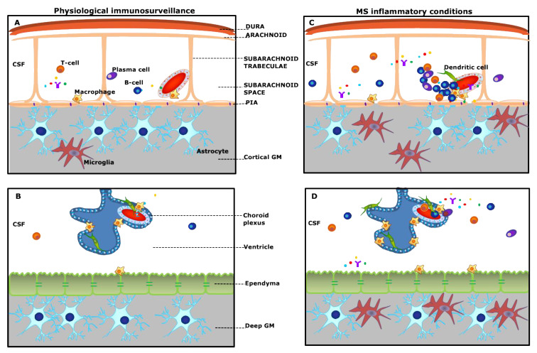
Progressive forms of multiple sclerosis (MS) are associated with chronic demyelination, axonal loss, neurodegeneration, cortical and deep gray matter damage, and atrophy. These changes are strictly associated with compartmentalized sustained inflammation within the brain parenchyma, the leptomeninges, and the cerebrospinal fluid. In progressive MS, molecular mechanisms underlying active demyelination differ from processes that drive neurodegeneration at cortical and subcortical locations. The widespread pattern of neurodegeneration is consistent with mechanisms associated with the inflammatory molecular load of the cerebrospinal fluid. This is at variance with gray matter demyelination that typically occurs at focal subpial sites, in the proximity of ectopic meningeal lymphoid follicles. Accordingly, it is possible that variations in the extent and location of neurodegeneration may be accounted for by individual differences in CSF flow, and by the composition of soluble inflammatory factors and their clearance. In addition, "double hit" damage may occur at sites allowing a bidirectional exchange between interstitial fluid and CSF, such as the Virchow-Robin spaces and the periventricular ependymal barrier. An important aspect of CSF inflammation and deep gray matter damage in MS involves dysfunction of the blood-cerebrospinal fluid barrier and inflammation in the choroid plexus. Here, we provide a comprehensive review on the role of intrathecal inflammation compartmentalized to CNS and non-neural tissues in progressive MS.

Keywords: autoimmunity; cerebrospinal fluid; choroid plexus; cytokines; demyelination; ependyma; inflammation; meninges; neurodegeneration.


Conflict of interest statement

The authors declare no conflict of interest.

Figures

Figure 1
Figure 1
Schematic overview of “meningeal and choroid plexus immunity” (A,B) and pathological alterations in multiple sclerosis (MS) (C,D). (A): The meninges consist of different membranes, including the dura mater (periosteal and meningeal layers), the arachnoid, and the pia. Strands of connective tissue, or arachnoid trabeculae, extend from the arachnoid layer and contains the vascular system. Under physiological conditions, the subarachnoid space is populated by scattered B- and T-cells, plasma cells, and macrophages and is filled with the cerebrospinal fluid (CSF) bathing the cortical grey matter (GM). (C): In MS, either innate or adaptive immune cells, in particular B-cells, accumulate in the subarachnoid space, outside the meningeal vessel, and may form ectopic tertiary lymphoid-like structures (TLSs), which are in close contact with the pia mater. This compartmentalized meningeal inflammation may contribute to the release in the CSF of inflammatory mediators that diffuse through the pia and induce subpial cortical demyelination. (B): The choroid plexus (CP) stroma is irrigated by fenestrated microvessels allowing free diffusion of blood-borne molecules. The CP epithelium surrounds the stroma and forms the blood–CSF barrier. Under physiological conditions, dendritic cells, macrophages, and sparse T-cells are found in the choroid plexus stroma. (D): During chronic neuroinflammation, both innate and adaptive immune cells accumulate in the choroid plexus and release inflammatory and proinflammatory factors in the surrounding CSF. These inflammatory changes may possibly induce changes in the structure and function of the ependyma lining the ventricle and the sub-ependymal glia, similar to those observed in the subpial GM.
Figure 2
Figure 2
Demyelinating cortical lesion and meningeal inflammation in chronic progressive MS. Immunohistochemistry for myelin oligodendrocyte protein (MOG) shows extensive subpial demyelination involving the neocortical grey matter, contiguous to MOG-positive brain tissue (A, black arrowheads). The lesion is in close proximity to inflamed meninges containing an elevated number of CD20+ B cells, either accumulated (B, arrow) or diffused (C), around subarachnoid vessels. Substantial increased density of MHC-class II+ activated microglia can be observed in the most external cortical layers (white arrowheads) at the pia/CSF boundary (D) nearby inflamed meninges. Original magnifications: 50× (A), 100× (D), 200× (B,C).
Figure 3
Figure 3
Periventricular demyelination and choroid plexus inflammation in chronic progressive MS. Immunohistochemistry for myelin oligodendrocyte protein (MOG) discloses subependymal demyelination (A) and an elevated number of MHC-class II+ activated microglia/macrophages (B) in the choroid plexus (arrows) and at the boundary ependyma/CSF (arrowheads). Elevated number of CD20+ B cells (C,E) and CD3+ T cells (D,F) are detected close to the tela choroidea (C,D), which separates the meninges (left) from the choroid plexus (right), and in the choroid plexus (E,F). Original magnifications: 100× (AF).

References

    1. Reich D.S., Lucchinetti C.F., Calabresi P.A. Multiple sclerosis. N. Engl. J. Med. 2018;378:169–180. doi: 10.1056/NEJMra1401483. - DOI - PMC - PubMed
    1. Brownlee W.J., Hardy T.A., Fazekas F., Miller D.H. Diagnosis of multiple sclerosis: Progress and challenges. Lancet. 2017;389:1336–1346. doi: 10.1016/S0140-6736(16)30959-X. - DOI - PubMed
    1. Absinta M., Lassmann H., Trapp B.D. Mechanisms underlying progression in multiple sclerosis. Curr. Opin. Neurol. 2020;33:277–285. doi: 10.1097/WCO.0000000000000818. - DOI - PMC - PubMed
    1. Baecher-Allan C., Kaskow B.J., Weiner H.L. Multiple sclerosis: Mechanisms and immunotherapy. Neuron. 2018;97:742–768. doi: 10.1016/j.neuron.2018.01.021. - DOI - PubMed
    1. Plum F. Ion Homeostasis in Cisternal and Lumbar Cerebrospinal Fluid. N. Engl. J. Med. 1975;293:1041–1042. doi: 10.1056/NEJM197511132932011. - DOI - PubMed

MeSH terms

LinkOut - more resources