. 2020 Nov 3;21(21):8223.
doi: 10.3390/ijms21218223.

Targeted Disruption of Mouse Dip2B Leads to Abnormal Lung Development and Prenatal Lethality

Affiliations

Targeted Disruption of Mouse Dip2B Leads to Abnormal Lung Development and Prenatal Lethality

Rajiv Kumar Sah et al. Int J Mol Sci. .

Abstract

Molecular and anatomical functions of mammalian Dip2 family members (Dip2A, Dip2B and Dip2C) during organogenesis are largely unknown. Here, we explored the indispensable role of Dip2B in mouse lung development. Using a LacZ reporter, we explored Dip2B expression during embryogenesis. This study shows that Dip2B expression is widely distributed in various neuronal, myocardial, endothelial, and epithelial cell types during embryogenesis. Target disruption of Dip2b leads to intrauterine growth restriction, defective lung formation and perinatal mortality. Dip2B is crucial for late lung maturation rather than early-branching morphogenesis. The morphological analysis shows that Dip2b loss leads to disrupted air sac formation, interstitium septation and increased cellularity. In BrdU incorporation assay, it is shown that Dip2b loss results in increased cell proliferation at the saccular stage of lung development. RNA-seq analysis reveals that 1431 genes are affected in Dip2b deficient lungs at E18.5 gestation age. Gene ontology analysis indicates cell cycle-related genes are upregulated and immune system related genes are downregulated. KEGG analysis identifies oxidative phosphorylation as the most overrepresented pathways along with the G2/M phase transition pathway. Loss of Dip2b de-represses the expression of alveolar type I and type II molecular markers. Altogether, the study demonstrates an important role of Dip2B in lung maturation and survival.

Keywords: Dip2b; LacZ expression; RNA sequencing; fetal lung development; growth restriction; mice; prenatal lethality.


Conflict of interest statement

The authors declare no conflict of interest. The funders had no role in the design of the study; in the collection, analyses, or interpretation of data; in the writing of the manuscript, or in the decision to publish the results.

Figures

Figure 1
Figure 1
Structure of the Dip2btm1a (KOMP) Wtsi allele: (A) Exons 8 and 9 are flanked by loxp while FRT, lacZ, neo, and FRT elements are inserted in intron 7. En2 SA sequences will trans-splice lacZ to exon 7 and be frame matched. LacZ-Neo-pA is a cDNA coding a fusion protein for both galactosidase as reporter and NEO expression as positive selection marker. PGK-DTA-pA is a negative selection marker for gene targeting in ES cells. KanR is a bacterial selection marker; (B) Genotyping by PCR. A 300 bp band appeared on gel for transgene allele and 350bp band for wild type allele. M, Molecular weight marker.
Figure 2
Figure 2
LacZ staining: (A) Whole mount of E9.5 Dip2btm1a/+ mice reveals lacZ expression in brain and dorsal tissues. Staining is intense at telencephalon (te), optic vesicle (ov), branchial arch (ba), limb bud (lb), and neural tube (nt); (B) At E11.5, Dip2b expression is concentrated in brain (mid brian, mb and hind brain, hb), dorsal neural tube (dsnt), intersomitic regions (so) and spinal cord (sc). At this stage, signals in heart and lung are also evident; (C) At E12.5, LacZ staining expands towards dorsal region specifically in brain (b), floor plate (fp) and whisker follicles (wf). Wild type littermates were devoid of lacZ staining; (D,E) Transverse sections of E15.5 and E18.5 Dip2btm1a/+ embryos were stained for LacZ and counter-stained with eosin. LacZ signals are shown in various neural tissues, in epithelial and endothelial cells of variety of organs. Olfactory epithelium (oe), mantle layer (ml), outflow tract (oft), blood vessels (bv), bronchus & bronchiole (br), pulmonary vessels (pv), epidermis (ep), and hair follicles (hf).
Figure 3
Figure 3
Phenotypical analysis: (A) Perinatal lethality in Dip2btm1a/tm1a mice. Survival plot show significant lethality of Dip2btm1a/tm1a mice at P0 (p < 0.0001, n = 36). Background picture shows cyanosis immediately after birth; (B) Representative images of newborns; (C) Body weight at E13.5—P0; (DF) Average weights (in grams) of brain, lung and heart at E18.5. Wet weights of organs were normalized to total body weight. * p < 0.05, **** p < 0.0001. NS, not significant.
Figure 4
Figure 4
Lung structure comparison of WT and Dip2btm1a/tm1a littermates: (A) Phenotype of a postnatal day 0 (P0) lungs after birth. The P0 lung was examined while it was floating in PBS; (B) The morphology of lung tissue and alveoli air space in WT and Dip2btm1a/tm1a at P0. H&E-stained (Scale bar, 75 μm) lungs is shown; (C) Number of alveolar air space in WT and and Dip2btm1a/tm1a mice; (D) Whole-mount of E11.5–E13.5 lungs of Dip2btm1a/tm1a (left) and wild-type (right) mice. CrL, right cranial lobe; ML, right medial lobe; CaL, right caudal lobe; AL, right accessory lobe; LL, left lobe; (E) The morphology of lung tissue in WT and Dip2btm1a/tm1a at E15.5. H&E-stained (Scale bar, 100 μm and 25 μm) lungs is shown. Col, columnar epithelium; Cub, cuboidal epithelium; (F) Quantification of distal epithelial buds in E15.5 lungs; (G,H) The morphology of lung tissue in WT and Dip2btm1a/tm1a at E17.5 and E18.5 is shown. Alveolar septation is shown by white arrow. H&E and DAPI stained (Scale bar, 75 μm and 25 μm) lungs are shown; (IK) Number of alveolar spaces, size and thickness in WT and Dip2btm1a/tm1a mice at E18.5; (L) Wet/dry ratio of E18.5 lung tissues. Data are means ± SD (n = 3, * p < 0.5, **** p < 0.0001 and NS: p = 0.11) calculated by standard two-tailed unpaired t-test.
Figure 5
Figure 5
Cell proliferation assay by Bromodeoxyuridine (BrdU) incorporation in lungs: (A) BrdU-Positive cell number in E15.5 lungs. Scale bars, 132.5 μm; (B) Cell proliferation assay at E18.5. Scale bars, 66.2 μm. NS = not significant, **** p < 0.0001 by student’s t-test. Three pubs were analyzed per group.
Figure 6
Figure 6
E18.5 Lung transcriptome analysis defined by FDR < 0.01 and Fold change > 2: (A) Volcano plots of differentially expressed genes (DEGs). The red and green dots represent up-regulated and downregulated genes respectively; (BE) Gene ontology (GO) analysis of DEGs; (B,C) Pie charts of top 10 overrepresented biological process terms from upregulated and downregulated genes respectively; (D,E) The mRNA expression levels of 11 & 10 genes annotated under biological process term ‘Cell cycle’ and ‘immune system process’ determined by qPCR. The red error bars represents standard deviation; (F,G) Pathway enrichment analysis; (F) KEGG pathway enrichment scatter plot. Each circle represents a KEGG pathway. The Y-axis represents the name of the pathway and the X-axis indicates Enrichment Factor, indicating the proportion of the annotated genes in the pathway. The lowest Q value represents the most significant pathway; (G) Biocarta G2/M phase transition pathway. A total of seven DEGs were annotated. Red arrow represents upregulated genes while green arrow represents downregulated genes.
Figure 7
Figure 7
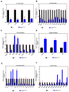
(AF) Gene expression analysis of alveolar, bronchiolar cell markers and lung mediator genes by qPCR of E18.5 lungs. Mix of total RNAs from three pups were analyzed per group. Data are means ± SD (n = 3, * p < 0.05 and ** p < 0.001).

References

    1. Tanaka M., Murakami K., Ozaki S., Imura Y., Tong X.-P., Watanabe T., Sawaki T., Kawanami T., Kawabata D., Fujii T., et al. DIP2 disco-interacting protein 2 homolog A (Drosophila) is a candidate receptor for follistatin-related protein/follistatin-like 1—Analysis of their binding with TGF-β superfamily proteins. FEBS J. 2010;277:4278–4289. doi: 10.1111/j.1742-4658.2010.07816.x. - DOI - PubMed
    1. Mukhopadhyay M., Pelka P., DeSousa D., Kablar B., Schindler A., Rudnicki M.A., Campos A.R. Cloning, genomic organization and expression pattern of a novel Drosophila gene, the disco-interacting protein 2 (dip2), and its murine homolog. Gene. 2002;293:59–65. doi: 10.1016/S0378-1119(02)00694-7. - DOI - PubMed
    1. Nitta Y., Yamazaki D., Sugie A., Hiroi M., Tabata T. DISCO Interacting Protein 2 regulates axonal bifurcation and guidance of Drosophila mushroom body neurons. Dev. Biol. 2017;421:233–244. doi: 10.1016/j.ydbio.2016.11.015. - DOI - PubMed
    1. Nitta Y., Sugie A. DISCO interacting protein 2 determines direction of axon projection under the regulation of c-Jun N-terminal kinase in the Drosophila mushroom body. Biochem. Biophys. Res. Commun. 2017;487:116–121. doi: 10.1016/j.bbrc.2017.04.028. - DOI - PubMed
    1. Noblett N., Wu Z., Ding Z.H., Park S., Roenspies T., Flibotte S., Chisholm A.D., Jin Y., Colavita A. DIP-2 suppresses ectopic neurite sprouting and axonal regeneration in mature neurons. J. Cell Biol. 2019;218:125–133. doi: 10.1083/jcb.201804207. - DOI - PMC - PubMed

LinkOut - more resources