

Automated 3D bio-imaging analysis of nuclear organization by NucleusJ 2.0
- PMID: 33153359
- PMCID: PMC7714466
- DOI: 10.1080/19491034.2020.1845012
Automated 3D bio-imaging analysis of nuclear organization by NucleusJ 2.0
Abstract
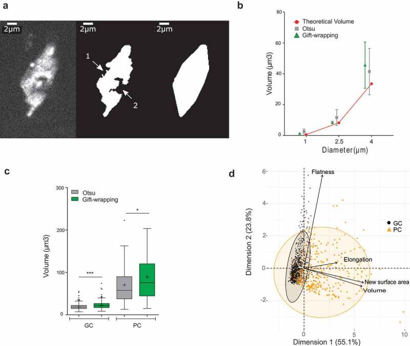
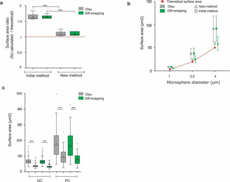
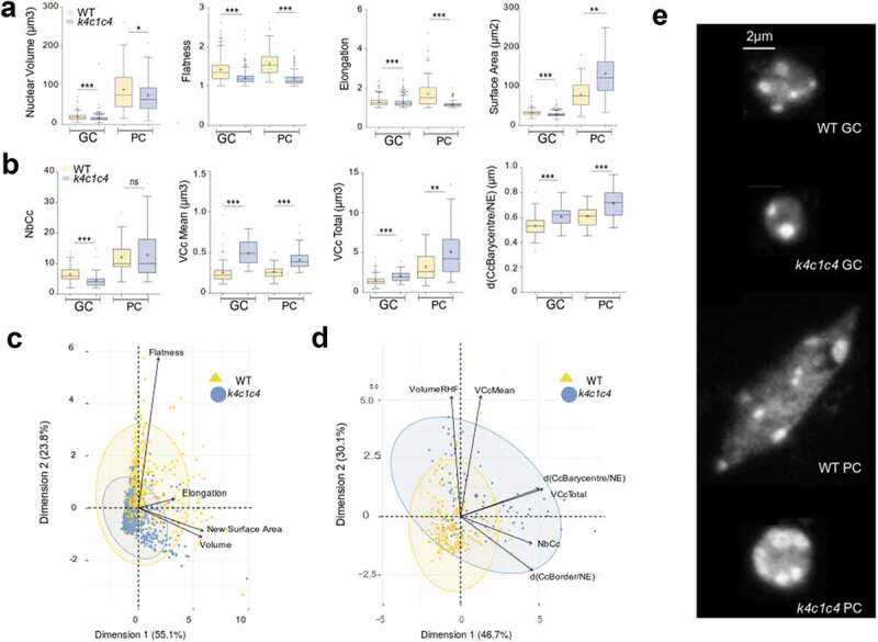
NucleusJ 1.0, an ImageJ plugin, is a useful tool to analyze nuclear morphology and chromatin organization in plant and animal cells. NucleusJ 2.0 is a new release of NucleusJ, in which image processing is achieved more quickly using a command-lineuser interface. Starting with large collection of 3D nuclei, segmentation can be performed by the previously developed Otsu-modified method or by a new 3D gift-wrapping method, taking better account of nuclear indentations and unstained nucleoli. These two complementary methods are compared for their accuracy by using three types of datasets available to the community at
Keywords: 3D DNA FISH; Three-dimensional microscopy imaging; image analysis; nuclear organization; plant nucleus.
Conflict of interest statement
No potential conflicts of interest were disclosed.
Figures
Similar articles
-
NucleusJ: an ImageJ plugin for quantifying 3D images of interphase nuclei.Bioinformatics. 2015 Apr 1;31(7):1144-6. doi: 10.1093/bioinformatics/btu774. Epub 2014 Nov 20. Bioinformatics. 2015. PMID: 25416749
-
NODeJ: an ImageJ plugin for 3D segmentation of nuclear objects.BMC Bioinformatics. 2022 Jun 6;23(1):216. doi: 10.1186/s12859-022-04743-6. BMC Bioinformatics. 2022. PMID: 35668354 Free PMC article.
-
Quantitative 3D Analysis of Nuclear Morphology and Heterochromatin Organization from Whole-Mount Plant Tissue Using NucleusJ.Methods Mol Biol. 2018;1675:615-632. doi: 10.1007/978-1-4939-7318-7_33. Methods Mol Biol. 2018. PMID: 29052214
-
Developing novel methods to image and visualize 3D genomes.Cell Biol Toxicol. 2018 Oct;34(5):367-380. doi: 10.1007/s10565-018-9427-z. Epub 2018 Mar 26. Cell Biol Toxicol. 2018. PMID: 29577183 Free PMC article. Review.
-
Quantitative analyses of the 3D nuclear landscape recorded with super-resolved fluorescence microscopy.Methods. 2017 Jul 1;123:33-46. doi: 10.1016/j.ymeth.2017.03.013. Epub 2017 Mar 18. Methods. 2017. PMID: 28323041 Review.
Cited by
-
The INDEPTH (Impact of Nuclear Domains on Gene Expression and Plant Traits) Academy: a community resource for plant science.J Exp Bot. 2022 Apr 5;73(7):1926-1933. doi: 10.1093/jxb/erac005. J Exp Bot. 2022. PMID: 35090020 Free PMC article.
-
Regulation of early seedling establishment and root development in Arabidopsis thaliana by light and carbohydrates.Planta. 2023 Sep 6;258(4):76. doi: 10.1007/s00425-023-04226-9. Planta. 2023. PMID: 37670114 Free PMC article.
-
Editorial for the SEB 2020 special issue 'dynamic organisation of the nucleus across kingdoms'.Nucleus. 2021 Dec;12(1):42-43. doi: 10.1080/19491034.2021.1883294. Nucleus. 2021. PMID: 33541203 Free PMC article. No abstract available.
-
Microfluidics to Follow Spatiotemporal Dynamics at the Nucleo-Cytoplasmic Interface During Plant Root Growth.Methods Mol Biol. 2025;2873:223-245. doi: 10.1007/978-1-0716-4228-3_13. Methods Mol Biol. 2025. PMID: 39576605
-
Deep learning -- promises for 3D nuclear imaging: a guide for biologists.J Cell Sci. 2022 Apr 1;135(7):jcs258986. doi: 10.1242/jcs.258986. Epub 2022 Apr 14. J Cell Sci. 2022. PMID: 35420128 Free PMC article. Review.
References
-
- Meier I, Richards EJ, Evans DE. Cell biology of the plant nucleus. Annu Rev Plant Biol. 2017;68:139–172. - PubMed
-
- Graumann K, Runions J, Evans DE. Characterization of SUN‐domain proteins at the higher plant nuclear envelope. Plant J. 2010;61:134–144. - PubMed
-
- Graumann K, Vanrobays E, Tutois S, et al. Characterization of two distinct subfamilies of SUN-domain proteins in Arabidopsis and their interactions with the novel KASH-domain protein AtTIK. J Exp Bot. 2014;65:6499–6512. - PubMed
-
- Tamura K, Iwabuchi K, Fukao Y, et al. Myosin XI-i links the nuclear membrane to the cytoskeleton to control nuclear movement and shape in Arabidopsis. Curr Biol. 2013;23:1776–1781. - PubMed
Publication types
MeSH terms
LinkOut - more resources
Full Text Sources
Other Literature Sources