. 2020 Nov 5;39(1):232.
doi: 10.1186/s13046-020-01690-z.

TM4SF1 promotes EMT and cancer stemness via the Wnt/β-catenin/SOX2 pathway in colorectal cancer

Affiliations

TM4SF1 promotes EMT and cancer stemness via the Wnt/β-catenin/SOX2 pathway in colorectal cancer

Qiang Tang et al. J Exp Clin Cancer Res. .

Abstract

Background: Transmembrane 4 L six family member 1 (TM4SF1) is upregulated in several epithelial cancers and is closely associated with poor prognosis. However, the role of TM4SF1 and its potential mechanism in colorectal cancer (CRC) remain elusive.

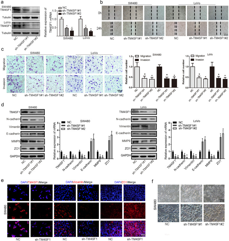
Methods: We investigated the expression of TM4SF1 in the Oncomine, the Cancer Genome Atlas (TCGA) and Gene Expression Omnibus (GEO) databases and confirmed the results by immunohistochemistry (IHC), qPCR and Western blotting (WB) of CRC tissues. The effect of TM4SF1 on the epithelial-to-mesenchymal transition (EMT) and cancer stemness of CRC cells was investigated by Transwell, wound healing and sphere formation assays. A series of in vitro and in vivo experiments were conducted to reveal the mechanisms by which TM4SF1 modulates EMT and cancer stemness in CRC.

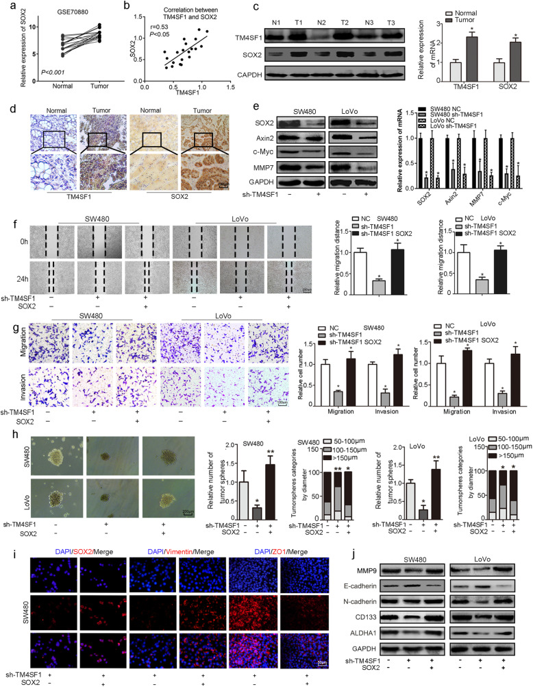
Results: TM4SF1 expression was markedly higher in CRC tissues than in non-tumour tissues and was positively correlated with poor prognosis. Downregulation of TM4SF1 inhibited the migration, invasion and tumour sphere formation of SW480 and LoVo cells. Conversely, TM4SF1 overexpression significantly enhanced the migration, invasion and tumoursphere formation potential of CRC cells, Additionally, TM4SF1 silencing inhibited the EMT mediated by transforming growth factor-β1 (TGF-β1). Mechanistically, gene set enrichment analysis (GSEA) predicted that the Wnt signalling pathway was one of the most impaired pathways in TM4SF1-deficient CRC cells compared to controls. The results were further validated by WB, which revealed that TM4SF1 modulated SOX2 expression in a Wnt/β-catenin activation-dependent manner. Furthermore, we found that knockdown of TM4SF1 suppressed the expression of c-Myc, leading to decreased c-Myc binding to the SOX2 gene promoter. Finally, depletion of TM4SF1 inhibited metastasis and tumour growth in a xenograft mouse model.

Conclusion: Our study substantiates a novel mechanism by which TM4SF1 maintains cancer cell stemness and EMT via the Wnt/β-catenin/c-Myc/SOX2 axis during the recurrence and metastasis of CRC.

Keywords: Colorectal cancer; EMT; SOX2; Stemness; TM4SF1; Wnt/β-catenin.


Conflict of interest statement

The authors declare no competing interests.

Figures

Fig. 1
Fig. 1
High TM4SF1 expression was significantly associated with poor prognosis. a, b Data from the Oncomine, TCGA and GEO databases showed that TM4SF1 was upregulated in CRC tissues compared to normal controls. c Immunohistochemical staining for TM4SF1 in CRC tissues and peritumoural normal tissues (n = 72). A total of 69% (50/72, SI ≥ 4) of CRC tissues were positive for TM4SF1 expression, while 31%(22/72, SI ≥3) of normal tissues were positive for TM4SF1. d, e High TM4SF1 expression showed a significant positive association with T stage and lymph node metastasis. f Kaplan-Meier survival analysis of TM4SF1 expression in the present study. g-i The overall survival curve was plotted by Kaplan-Meier Plotter in the R2 Genomics Analysis Platform (https://hgserver1.amc.nl/cgi-bin/r2/main.cgi; the study conducted by Sveen, Smith and Marisa). j-k qRT-PCR and WB analysis revealed that TM4SF1 was upregulated in CRC tissues (*P < 0.05). l WB and qPCR analysis of TM4SF1 expression in CRC cell lines (HCT116, SW480, DLD1, LoVo, RKO) and normal colon mucosal epithelial cells (NCM460 and FHC) *P < 0.05
Fig. 2
Fig. 2
TM4SF1 deficiency suppresses the migration and invasion of CRC cells. a WB and qRT-PCR analysis of the efficiency of sh-TM4SF1 and sh-Control (NC) transfection in SW480 and LoVo cells. b Wound healing assays of cell migration in SW480 and LoVo cells. The images of wound closure are presented at the indicated number of hours after scratching (0, 24 h). c Transwell assays were performed to examine the potential migration and invasion of TM4SF1 KD cells or negative control cells. d WB and qRT-PCR analysis of the expression of EMT markers in CRC cells transfected with sh-TM4SF1. e Immunofluorescence staining showed the changes in the expression of EMT-associated genes vimentin and ZO1 (red) in SW480 cells. Nuclei were counterstained with DAPI (blue). f Morphological change of SW480 cells transfected with sh-TM4SF1 and NC. *P < 0.05
Fig. 3
Fig. 3
TM4SF1 is involved in TGF-β1-induced EMT, and modulates the stemness properties. a Wound healing assays showed that TGF-β1 enhanced the migration potential. TM4SF1 silencing decreased the migration of TGF-β1-treated SW480 cells. b Transwell assays showed that TGF-β1 enhanced the migration and invasion potential. TM4SF1 silencing suppressed the migration and invasion of TGF-β1-treated SW480 cells. c WB analysis showed that TGF-β1 increased the expression of Smad2, and EMT markers (vimentin, N-cadherin, MMP9). d TM4SF1 knockdown downregulated the expression of EMT markers in TGF-β1-treated LoVo cells. e Tumoursphere assay of CRC cells transfected with sh-TM4SF1 and NC. f CCK-8 assay showed that CRC cells with sh-TM4SF1 showed suppressed survival after treatment with fluorouracil. g. WB analysis of stemness markers (SOX2, CD44, CD133, ALDHA1) in CRC cells transfected with sh-TM4SF1. *P < 0.05
Fig. 4
Fig. 4
TM4SF1 drives the EMT and cancer stemness of CRC through Wnt/β-catenin signalling. a GSEA indicated several dysregulated Kyoto Encyclopedia of Genes and Genomes (KEGG) pathways. b Heat map of the indicated target genes in TM4SF1-silenced and control cells. The target genes of Wnt/β-catenin signalling were decreased in TM4SF1-silenced cells. c TM4SF1 knockdown reduced the levels of total β-catenin and the nuclear translocation of β-catenin. d, e LiCl restored the expression of Wnt/β-catenin target genes, EMT and stemness markers in TM4SF1-deficient cells. f-g Activation of β-catenin rescued the EMT phenotype and diminished the TM4SF1 deficiency-mediated inhibitory effect on cell migration and invasion. h LiCl promoted tumour sphere formation in TM4SF1-deficient cells. The tumourspheres were counted, and the percentage of tumourspheres with diameters 50–100 μm, 100–150 μm, and > 150 μm was calculated. *P < 0.05
Fig. 5
Fig. 5
TM4SF1 promotes the EMT of CRC cells via the Wnt/β-catenin/SOX2 signalling pathway. a The GEO database (GSE70880) indicated that SOX2 was overexpressed in CRC tissues. b Spearman’s rank correlation analysis showed the correlation between the expression of TM4SF1 and SOX2 in CRC tissues. c WB and qRT-PCR analysis showed the expression of TM4SF1 and SOX2 in CRC tissues and adjacent non-tumour tissues. d Immunohistochemical staining for TM4SF1 and SOX2 in CRC tissues and normal tissues. e TM4SF1 silencing downregulated the expression of SOX2, Axin2, MMP7, and c-Myc at the protein and mRNA levels. f Wound healing assays showed that SOX2 overexpression rescued the inhibitory effects of migration of sh-TM4SF1-SW480/LoVo cells. g Transwell assays showed that SOX2 overexpression enhanced the migration and invasion potential of TM4SF1-silenced cells. h Tumoursphere assay of CRC cells transfected with sh-TM4SF1 cells or SOX2 cDNA. The tumoursphere number was counted, and the percentage of tumourspheres with diameters < 100 μm, 100–150 μm or > 150 μm was calculated and plotted. i Immunofluorescence images of SOX2, vimentin and ZO1 (red) with SOX2 overexpression in TM4SF1-deficient cells. j Upregulation of SOX2 expression attenuated the loss of expression of EMT and stemness markers after TM4SF1 silencing. *P < 0.05
Fig. 6
Fig. 6
c-Myc directly binds the SOX2 promoter and regulates SOX2 expression. a LiCl significantly upregulated the expression of SOX2 and c-Myc in TM4SF1 knockdown CRC cells. b SOX2 deficiency significantly downregulated the expression of SOX2 but not the expression of c-Myc in the LiCl-treated cells. c c-Myc knockdown decreased the expression of SOX2. d c-Myc overexpression upregulated the expression of SOX2. e Promoter region of the SOX2 gene with a putative c-Myc binding site. f ChIP assays assessing the binding of c-Myc to the SOX2 promoter. Normal rabbit IgG and anti-c-Myc antibody were used to precipitate chromatin DNA fragments as indicated. g, h SOX2 knockdown suppressed migration and tumour sphere formation in c-Myc-overexpressing cells. i SOX2 deficiency decreased the expression of N-cadherin, CD133, and CD44 and upregulated the expression of E-cadherin in c-Myc-overexpressing cells. *P < 0.05
Fig. 7
Fig. 7
TM4SF1 promotes tumourigenesis and tumour metastasis in mice. a, b Xenograft weight (mg) and size (cm) were measured. c Immunohistochemistry staining of TM4SF1, SOX2, β-catenin, CD133 and vimentin in xenograft tissues. d WB analysis of the expression of TM4SF1, SOX2, and β-catenin expression from sh-TM4SF1/scramble xenograft tumours. e Abolished tumour formation was found in the lungs of SW480 cell-injected mice. The statistical data of the tumour foci number and diameter are presented, and the black arrow marked metastatic nodules. H&E stained histological images of the lungs of the two groups of mice. *P < 0.05

References

    1. Ferlay J, Soerjomataram I, Dikshit R, Eser S, Mathers C, Rebelo M, et al. Cancer incidence and mortality worldwide: sources, methods and major patterns in GLOBOCAN 2012. Int J Cancer. 2015;136(5):E359–EE86. doi: 10.1002/ijc.29210. - DOI - PubMed
    1. Song M, Garrett WS, Chan AT. Nutrients, foods, and colorectal cancer prevention. Gastroenterology. 2015;148(6):1244–60e16. - PMC - PubMed
    1. Mazzocca A, Carloni V, Sciammetta SC, Cordella C, Pinzani M. Expression of transmembrane 4 superfamily (TM4SF) proteins and their role in hepatic stellate cell motility and wound healing migration. J Hepatol. 2002;37(3):322–330. doi: 10.1016/S0168-8278(02)00175-7. - DOI - PubMed
    1. Lin C-I, Merley A, Sciuto TE, Li D, Dvorak AM, Melero-Martin JM, et al. TM4SF1: a new vascular therapeutic target in cancer. Angiogenesis. 2014;17(4):897–907. doi: 10.1007/s10456-014-9437-2. - DOI - PMC - PubMed
    1. Hellstrom IHD, Linsley P, Brown JP, Brankovan V, Hellstrom KE. Monoclonal mouse antibodies raised against human lung carcinoma. Elsevier. 1987;3(1):3917–23. - PubMed

MeSH terms