. 2021 Jan 1;371(6524):eabc5667.
doi: 10.1126/science.abc5667. Epub 2020 Nov 5.

Structural basis of antagonizing the vitamin K catalytic cycle for anticoagulation

Affiliations

Structural basis of antagonizing the vitamin K catalytic cycle for anticoagulation

Shixuan Liu et al. Science. .

Abstract

Vitamin K antagonists are widely used anticoagulants that target vitamin K epoxide reductases (VKOR), a family of integral membrane enzymes. To elucidate their catalytic cycle and inhibitory mechanism, we report 11 x-ray crystal structures of human VKOR and pufferfish VKOR-like, with substrates and antagonists in different redox states. Substrates entering the active site in a partially oxidized state form cysteine adducts that induce an open-to-closed conformational change, triggering reduction. Binding and catalysis are facilitated by hydrogen-bonding interactions in a hydrophobic pocket. The antagonists bind specifically to the same hydrogen-bonding residues and induce a similar closed conformation. Thus, vitamin K antagonists act through mimicking the key interactions and conformational changes required for the VKOR catalytic cycle.


Figures

Figure 1.
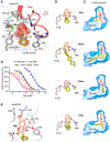
Figure 1.. Overall structure of HsVKOR with bound warfarin.
(A) Warfarin inhibition of epoxide reductase activity in cultured cells. The inhibition curves of untagged and sfGFP-fused HsVKOR and TrVKORL are compared. The activity assay has been repeated three times. (B) Structure of HsVKOR (side view) with warfarin (War). Secondary structure elements in the large ER luminal domain are named and presented in different colors. The four transmembrane helices (TM1–4) are shown in grey. The 4-hydroxycoumarin and side groups of warfarin are colored in orange and yellow green, respectively. The overall structure of HsVKOR with warfarin and with other vitamin K antagonists are very similar (fig. S3E). (C) Surface representation of the structure (exposed view). Warfarin is bound at the central pocket that contains the active site cysteines. The cap helix forms top part of the central pocket. The tunnel below is expected to bind the isoprenyl chain of K or KO substrate. (D) Top view of the structure.
Figure 2.
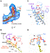
Figure 2.. Critical molecular interactions between HsVKOR and vitamin K antagonists.
(A) The warfarin-binding pocket. The Fo-Fc omit map of warfarin is contoured at 3σ (red mesh) and −3σ (blue mesh). Hydrogen bonding interactions are indicated by dashed green lines. (B) Similar hydrogen-bonding patterns are observed for different VKAs (Brod: brodifacoum, Phen: phenindione, Chlo: chlorophacinone). Standard atomic numbers for the key positions of VKAs are indicated. (C) Side groups (yellow green) of VKAs bind differently to the isoprenyl-chain tunnel (surface view). (D) Inhibition curves of VKAs and hydroxywarfarins against HsVKOR using the cell-based assay. (E) Models of the 6- and 7-hydoxywarfarin in the warfarin-binding pocket. The dashed green line indicates a putative hydrogen bond to the 6-hydroxyl group, and the red curve represents a lack of favorable interaction between the polar 7-hydroxyl group and the hydrophobic protein side chains.
Figure 3.
Figure 3.. TrVKORL structures showing warfarin-induced conformational changes.
(A) Overall structure of TrVKORL with warfarin (colored by structure elements) is nearly the same as that of HsVKOR with warfarin (blue). (B) Similarity of their warfarin binding interactions. TrVKORL residues are colored in grey (carbon atoms) and HsVKOR residues in blue. The Fo-Fc omit map of warfarin is contoured at 3σ (red mash) and −3σ (blue mesh). C) TrVKORL with bound warfarin adopts a closed conformation, in which the cap helix and β-hairpin are formed and interact with loop 3–4. (D) The ligand-free TrVKORL adopts an open conformation. In the luminal helix, residues converted from the loop 1 and β-hairpin (in C) are colored in blue and purple, respectively. The TMs are colored in dark grey to indicate the open conformation. (E-F) Detailed view of the closed (E) and open (F) states. Warfarin binding induces the movements of Cys43-Cys51, Val54, and Phe55 and promotes the stabilizing interactions of Asp44 (dashed lines). See Movie S2 for modeled structural transition.
Figure 4.
Figure 4.. Structures of vitamin K-bound TrVKORL in noncatalytic and catalytic states.
(A) Fully oxidized TrVKORL with K. The central pocket, including the isoprenyl-chain tunnel, are shown in surface view. The Fo-Fc omit map of K is contoured at 3σ (red mash), −3σ (no visible density), and 2.5 σ (blue mash) due to the weak density of K in this redox state. The distance between the C2 atom of K and the Cys135 sulfur is indicated. (B) TrVKORL Cys132Ser mutant with K. The Fo-Fc omit map of K is contoured at 3σ (red mash) and −3σ (light blue mesh). The reduced Cys135 and K form a charge-transfer complex (dashed arrow). (C) Open conformation in the fully oxidized state. (D) Closed conformation with formation of the charge-transfer complex. The unimpeded electron-transfer path between the four cysteines and K is highlighted by the orange shading. (E) Superimposed structures of TrVKORL in the K-bound (as in (D); colored by structural elements) and warfarin-bound (as in Fig. 3C; blue) states. (F) Similar hydrogen-bonding interactions with K (orange and yellow green) and with warfarin (blue).
Figure 5.
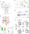
Figure 5.. Structure of HsVKOR bound to a KO adduct.
(A) Overall structure of HsVKOR Cys43Ser mutant co-crystallized with KO. The dashed line indicates the disordered region between TM1e and cap domain. (B) Detailed view of the active site. The Fo-Fc omit map of C135-KOH is contoured at 3σ (red mash) and −3σ (blue mesh) ; 3-OH K is modeled into the density based on the geometric restraint and the catalytic chemistry (26, 27). The short distance between the C2 atom of 3-OH K and the sulfhydryl group of Cys135 suggests that they are connected by a C–S bond. (C) Two-dimensional 1H-13C HSQC spectrum (full spectrums in fig. S13) showing signals of 13C-labeled 2-methyl groups in four products of KO reduction with dithiothreitol (DTT) (52). Chemical structures of the products and the assigned chemical shifts are indicated. The chemical shifts of 13C 2-methyl group are strongly influenced by the ring current field of the naphthoquinone group (the 2-methyl is in plane with the naphthoquinone ring for K, in tetrahedral angle for 3-OH K and in other angles for KO and mercapto 3-OH K). (D) Superimposed regions of three 2D 1H-13C HSQC spectra (full spectra in fig. S13-S15) showing mercapto reduction products of 2-methyl-13C-labeled KO. Signals of products recovered from TrVKORL Cys43Ser catalyzed reaction are shown in blue and those from Cys135Ser in red. The non-enzymatic (DTT-reduced) products signals are shown in black. (E) Superimposed spectra (full spectra in fig. S16-S18) of reaction products obtained from wild-type HsVKOR (WT; green), Asn80Ala (blue) and Tyr139Phe mutants (orange) with KO and DTT. An area of 2D HSQC spectrum containing 1H-13C peaks of 2-methyl-13C-labeled K (2.20-15.5 ppm), KO (1.77-14.7 ppm) and 3-OH K (1.50-10.6 ppm) is shown. Inset, relative KO reduction activity of HsVKOR constructs in microsomes (see Materials and Methods). (F) The catalytic cycle and inhibition of HsVKOR is accompanied with redox-state and conformation changes. The large luminal domain is shown as a hemisphere (pink) and the transmembrane domain as a cylinder (light grey: closed conformation, dark grey: open conformation). State I, The partially oxidized state with free Cys43 and free Cys135. State II, Cys135 forms a stable adduct with 3-OH K (KOH) or K (KH), whose binding induces the closed conformation and the juxtaposition of Cys43 for electron transfer (dashed arrow). State III, The reduced Cys132 attacks Cys135–K-OH (or KH) to generate K (or KH2). State IV, The fully oxidized state is in an open conformation to release K (or KH2). Left, Warfarin (W) competes with the substrates for the partially oxidized enzyme. Unlike the substrates, warfarin binds also to the fully oxidized enzyme and removes it from the enzyme pool. The bound warfarin locks HsVKOR in both of the redox states into a closed conformation.

Similar articles

Cited by

References

    1. Goy J, Crowther M, Approaches to diagnosing and managing anticoagulant-related bleeding. Seminars in Thrombosis and Hemostasis 38, 702–710 (2012). - PubMed
    1. Schulman S, Beyth RJ, Kearon C, Levine MN, Hemorrhagic complications of anticoagulant and thrombolytic treatment: American College of Chest Physicians evidence-based clinical practice guidelines (8th edition). (2008), vol. 133. - PubMed
    1. Budnitz DS, Lovegrove MC, Shehab N, Richards CL, Emergency hospitalizations for adverse drug events in older Americans. The New England journal of medicine 365, 2002–2012 (2011). - PubMed
    1. Stafford DW, The vitamin K cycle. J Thromb Haemost 3, 1873–1878 (2005). - PubMed
    1. Li W et al., Structure of a bacterial homologue of vitamin K epoxide reductase. Nature 463, 507–512 (2010). - PMC - PubMed

Publication types