. 2020 Dec 4;370(6521):1208-1214.
doi: 10.1126/science.abe0075. Epub 2020 Nov 5.

De novo design of potent and resilient hACE2 decoys to neutralize SARS-CoV-2

Affiliations

De novo design of potent and resilient hACE2 decoys to neutralize SARS-CoV-2

Thomas W Linsky et al. Science. .

Abstract

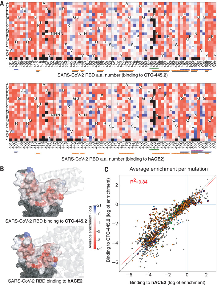
We developed a de novo protein design strategy to swiftly engineer decoys for neutralizing pathogens that exploit extracellular host proteins to infect the cell. Our pipeline allowed the design, validation, and optimization of de novo human angiotensin-converting enzyme 2 (hACE2) decoys to neutralize severe acute respiratory syndrome coronavirus 2 (SARS-CoV-2). The best monovalent decoy, CTC-445.2, bound with low nanomolar affinity and high specificity to the receptor-binding domain (RBD) of the spike protein. Cryo-electron microscopy (cryo-EM) showed that the design is accurate and can simultaneously bind to all three RBDs of a single spike protein. Because the decoy replicates the spike protein target interface in hACE2, it is intrinsically resilient to viral mutational escape. A bivalent decoy, CTC-445.2d, showed ~10-fold improvement in binding. CTC-445.2d potently neutralized SARS-CoV-2 infection of cells in vitro, and a single intranasal prophylactic dose of decoy protected Syrian hamsters from a subsequent lethal SARS-CoV-2 challenge.


Figures

Fig. 1
Fig. 1. Design and characterization of de novo ACE2 decoys.
(A) ACE2 (gray) and its binding motifs (H1 19-52, orange; H2 55-84, green; EE3 346-360, blue) in complex with SARS-CoV-2 RBD (pink). Three starting structures were simultaneously used as targets (see main text); 6VW1 is shown. (B) De novo secondary structure elements (magenta) were computationally generated to stabilize H1, H2, and EE3. Seven combinations of secondary structure elements were considered. Circles are α-helices, triangles are β-sheets, filled circles are helices oriented forward, and empty circles are helices oriented backward. We used Rosetta to generate fully connected backbones (using the “protein_mimic_designer” algorithm) and amino acid sequences predicted to fold into the target structure. In all cases, the binding interface of ACE2 with the SARS-CoV-2 RBD was preserved intact (see the materials and methods). (C) Automatic computational filtering based on eight metrics selected the best candidates. The RMSD of the binding motifs to ACE2 was also used as a quality check. The dots indicate the mean computational score for each design scored against the three target RBD structures. Designs selected for experimental testing are shown in black. Our best design, CTC-445, is shown in red. The blue boxes indicate the filtering thresholds (see the materials and methods). (D) Designs that passed filtering were subjected to biased forward folding simulations (see the materials and methods), here shown for CTC-445, including the unsalted biased simulation (brown), the native-salted simulation (orange), and relaxation (blue). (E) The top 196 designs were selected for yeast display screening using a combination of Rosetta score per residue, the ddG Rosetta filter, and the folding simulations (see the materials and methods). The designs were individually assessed for specific binding to SARS-CoV-2 spike RBD (Fc fusion, 200 nM). The plot for CTC-445 is shown. (F) CTC-445 was recombinantly expressed and purified by affinity chromatography (see the materials and methods). Analytical size exclusion chromatography (SEC) for CTC-445 revealed the presence of oligomeric species. (G and H) CTC-445 was optimized by directed evolution and rational combination of the observed favorable mutations (G), leading to CTC-445.2 (SEC), which is mainly monomeric in solution (H) and ~1000× more potent to compete with ACE2 than its parent [see (G)]. We further optimized the potency of our molecule by generating a bivalent version named CTC-445.2d. (I) Potency of designs to outcompete binding of SARS-CoV-2 RBD to ACE2, as measured by competition enzyme-linked immunosorbent assay (ELISA) using a constant concentration of 0.4 nM ACE2. (J) Timeline of the de novo protein design and optimization pipeline. Timewise, green indicates phases that we believe were performed optimally, red indicates those that can potentially be avoided in future efforts, and yellow indicates phases that can potentially be expedited by using more advanced and/or automated methods for gene synthesis, cloning, and high-throughput screening.
Fig. 2
Fig. 2. Stability and binding of the de novo protein decoys CTC-445, CTC-445.2, and CTC-445.2d.
(A) Design models of CTC-445, CTC-445.2, and CTC-445.2d. CTC-445.2 contains five mutations that were guided by directed evolution experiments. CTC-445.2d is a bivalent variant composed of two CTC-445.2 subunits linked by a 16-mer flexible GS linker (sequence -GGGSGGSGSGGSGGGS-). (B) Circular dichroism of recombinantly expressed CTC-445 (red), CTC-445.2 (blue), and CTC-445.2d (orange). Thermally induced melting of the decoys was followed by its circular dichroism signal at 208 nm (heating rate, 2°C/min). The inset shows far ultraviolet (UV) wavelength spectra at 20°C (purple), after heating to ~95°C (brown), and after cooling the heated sample to 20°C (green dashed). Complete ellipticity spectra recovery (full reversibility) upon cooling was observed in all cases. Calculated Tm values for CTC-445, CTC-445.2, and CTC-445.2d are 75.3 ± 0.2°C, ≈93°C, and 71.7.± 0.2°C, respectively. (C) Binding was assessed using biolayer interferometry (OCTET) binding assays of CTC-445, CTC-445.2, and CTC-445.2d against immobilized SARS-CoV-2 RBD (top) or SARS-CoV-1 RBD (bottom) (see table S1). The model fitting is shown with dotted black lines.
Fig. 3
Fig. 3. Cryo-EM structure of the CTC-445.2–S complex.
(A to D) Cryo-EM reconstructions of CTC-445.2 (blue) bound to soluble spike trimers (gray). 3D classification revealed four distinct classes: one CTC-445.2 bound to an “up” RBD (A), two CTC-445.2 bound to two “up” RBDs (B), two CTC-445.2 bound to one “up” and one “down” RBD (C), and three CTC-445.2 bound to two “up” and one “down” RBD (D). (E) Overlay of CTC-445.2-RBD computationally modeled (yellow) and experimentally determined using cryo-EM (blue). The Cα RMSD between the design model and the refined experimental structure is 1.1 Å. (F to H) Comparison of cryo-EM CTC-445.2 (blue), computationally modeled CTC-445.2 (yellow), and hACE2 (green) at the interface of the RBD (gray). (I) Deep mutational scanning heatmap showing the average effect on the enrichment for single site mutants of CTC-445.2 when assayed by yeast display for binding to the SARS-CoV-2 RBD (binding assayed at RBD concentrations of 100, 50, 25, 12.5, 6.25, 3.125, and 1.5625 pM; see the materials and methods). (J) Design model of CTC-445.2 colored by average enrichment at each residue position [from the data in (I)] bound to SARS-CoV-2 RBD (gray). As expected, mutations in the core of the design or to positions involved in binding to the RBD are generally disallowed. The deep mutational scanning revealed that there is still room to further improve the binding affinity of CTC-445.2, including mutations in the binding interface that in principle could afford higher potency and selectivity at the cost of compromising the decoy’s mutational escape resiliency (see Fig. 4).
Fig. 4
Fig. 4. Resilience of CTC-445.2 to SARS-CoV-2 RBD mutational escape.
(A) Deep mutational scanning (DMS) of the SARS-CoV-2 RBD interface was performed to assess the effect on binding (by yeast display) to CTC-445.2 (top) or hACE2 (bottom) at eight different concentrations (656, 218, 72, 24, 8, 2, 0.3, and 0.1 nM; fig. S16 and materials and methods). The heatmaps indicate the effect on binding for each possible single amino acid mutation in the hACE2-binding interface of the RBD (see the materials and methods). The results are the average over all the concentrations tested. A black square represents lack of expression in the naive (unselected) library. The color bars at the bottom indicate the secondary structure element with which a given RBD residue interacts: H1, orange; H2, green; EE3, blue; and H4, magenta. Approximately 1700 single mutations were targeted by the experiment. (B) The SARS-CoV-2 RBD surface is colored according to the per-residue-averaged enrichments for binding to CTC-445.2 (top) or hACE2 (bottom). For reference, the structure of CTC-445.2 or ACE2 (respectively) is shown in semitransparent gray cartoons. (C) The 2D scatter plots compare the enrichment values [as in (A)] for the DMS of the RBD binding to CTC-445.2 (y-axis) versus hACE2 (x-axis). There is a high correlation between the effect of RBD mutations in the binding of both molecules, demonstrating the mutational resilience of the de novo decoy (Pearson’s r = 0.92).
Fig. 5
Fig. 5. In vitro virus neutralization by CTC-445.2d.
(A) Top left: In vitro neutralization of NanoLuc SARS-CoV-2 by CTC-445.2d in Calu-3 cells after 72 hours of incubation at a MOI of 1.0. Top right: A cell viability assay (48 hours) confirmed that the decoys are not cytotoxic to Calu-3. Bottom left: In vitro neutralization of live BetaCoV/Hong Kong/VM20001061/2020 SARS-CoV-2 virus in Vero E6 cells at a MOI of 1.0. The cells were incubated with CTC-445.2d throughout infection and the colors indicate the following: orange, before infection, during infection, and after infection; black, after infection only; and gray, before infection only. SARS-CoV-2 RNA copy numbers were determined by quantitative real-time reverse transcription polymerase chain reaction. All assays were performed in triplicate unless otherwise noted, and all data points are shown. Bottom right: Cell viability in Vero E6 cells was independently performed (CCK8 assay) and it was confirmed that the de novo decoys are not cytotoxic. (B) In vivo mouse pharmacokinetics and tolerability of intranasally administered CTC-445.2d. Left: Plot showing the concentration of fully functional CTC-445.2d (i.e., capable of binding to the SARS-CoV-2 RBD; see the materials and methods) found in homogenized lungs of Balb/c mice after a single 100 μg dose, measured at various times after dosing (n = 5 mice). Right: Body weight of mice after repeat daily intranasal doses of CTC-445.2d (100 μg; n = 18 at day 0) compared with control [phosphate-buffered saline (PBS)–treated] mice (n = 5). At each time point, three CTC-445.2d–treated mice were sacrificed for lung examination. Weight data shown are for the remaining mice (n = 18, 15, 12, 9, 6, and 3 at days 1, 2, 4, 8, 11, and 14, respectively). No significant weight loss or lung abnormalities were observed. Error bars indicate the standard deviation. (C) In vivo Syrian hamster SARS-CoV-2 challenge. Left: Body weight measurements through day 10 for unchallenged hamsters (n = 5, red) compared with SARS-CoV-2–challenged hamsters treated either with a single dose of CTC-445.2d (day 0 at –12 hours; n = 8, orange) or PBS (day –1, day 0 at –12 hours, day 1, and day 2; n = 7, gray). Right: Survival plot. Hamsters were euthanized when they displayed clinical signs of distress according to protocol clinical scoring criteria (see the materials and methods). At the end of the experiment, all hamsters treated with the de novo decoy CTC-445.2d survived, exhibiting moderate weight loss, whereas hamsters treated with vehicle did not survive past day 7 because of severe weight loss and other complications from the viral infection (see table S5).

Update of

References

    1. Gordon D. E., Jang G. M., Bouhaddou M., Xu J., Obernier K., White K. M., O’Meara M. J., Rezelj V. V., Guo J. Z., Swaney D. L., Tummino T. A., Hüttenhain R., Kaake R. M., Richards A. L., Tutuncuoglu B., Foussard H., Batra J., Haas K., Modak M., Kim M., Haas P., Polacco B. J., Braberg H., Fabius J. M., Eckhardt M., Soucheray M., Bennett M. J., Cakir M., McGregor M. J., Li Q., Meyer B., Roesch F., Vallet T., Mac Kain A., Miorin L., Moreno E., Naing Z. Z. C., Zhou Y., Peng S., Shi Y., Zhang Z., Shen W., Kirby I. T., Melnyk J. E., Chorba J. S., Lou K., Dai S. A., Barrio-Hernandez I., Memon D., Hernandez-Armenta C., Lyu J., Mathy C. J. P., Perica T., Pilla K. B., Ganesan S. J., Saltzberg D. J., Rakesh R., Liu X., Rosenthal S. B., Calviello L., Venkataramanan S., Liboy-Lugo J., Lin Y., Huang X.-P., Liu Y., Wankowicz S. A., Bohn M., Safari M., Ugur F. S., Koh C., Savar N. S., Tran Q. D., Shengjuler D., Fletcher S. J., O’Neal M. C., Cai Y., Chang J. C. J., Broadhurst D. J., Klippsten S., Sharp P. P., Wenzell N. A., Kuzuoglu-Ozturk D., Wang H.-Y., Trenker R., Young J. M., Cavero D. A., Hiatt J., Roth T. L., Rathore U., Subramanian A., Noack J., Hubert M., Stroud R. M., Frankel A. D., Rosenberg O. S., Verba K. A., Agard D. A., Ott M., Emerman M., Jura N., von Zastrow M., Verdin E., Ashworth A., Schwartz O., d’Enfert C., Mukherjee S., Jacobson M., Malik H. S., Fujimori D. G., Ideker T., Craik C. S., Floor S. N., Fraser J. S., Gross J. D., Sali A., Roth B. L., Ruggero D., Taunton J., Kortemme T., Beltrao P., Vignuzzi M., García-Sastre A., Shokat K. M., Shoichet B. K., Krogan N. J., A SARS-CoV-2 protein interaction map reveals targets for drug repurposing. Nature 583, 459–468 (2020). 10.1038/s41586-020-2286-9 - DOI - PMC - PubMed
    1. Liu C., Zhou Q., Li Y., Garner L. V., Watkins S. P., Carter L. J., Smoot J., Gregg A. C., Daniels A. D., Jervey S., Albaiu D., Research and development on therapeutic agents and vaccines for COVID-19 and related human coronavirus diseases. ACS Cent. Sci. 6, 315–331 (2020). 10.1021/acscentsci.0c00272 - DOI - PMC - PubMed
    1. Chen W.-H., Strych U., Hotez P. J., Bottazzi M. E., The SARS-CoV-2 vaccine pipeline: An overview. Curr. Trop. Med. Rep. 7, 1–4 (2020). 10.1007/s40475-020-00201-6 - DOI - PMC - PubMed
    1. Chan K. K., Dorosky D., Sharma P., Abbasi S. A., Dye J. M., Kranz D. M., Herbert A. S., Procko E., Engineering human ACE2 to optimize binding to the spike protein of SARS coronavirus 2. Science 369, 1261–1265 (2020). 10.1126/science.abc0870 - DOI - PMC - PubMed
    1. J. D. Walter, C. A. J. Hutter, I. Zimmermann, J. Earp, P. Egloff, M. Sorgenfrei, L. M. Hürlimann, I. Gonda, G. Meier, S. Remm, S. Thavarasah, P. Plattet, M. A. Seeger, Sybodies targeting the SARS-CoV-2 receptor-binding domain. bioRxiv 045419 [Preprint]. 16 May 2020. 10.1101/2020.04.16.045419. - DOI

Publication types

MeSH terms