Review
. 2021 Jan-Jun:296:100035.
doi: 10.1074/jbc.REV120.014915. Epub 2020 Dec 1.

Stem cell fate determination through protein O-GlcNAcylation

Affiliations
Review

Stem cell fate determination through protein O-GlcNAcylation

Muhammad Abid Sheikh et al. J Biol Chem. 2021 Jan-Jun.

Abstract

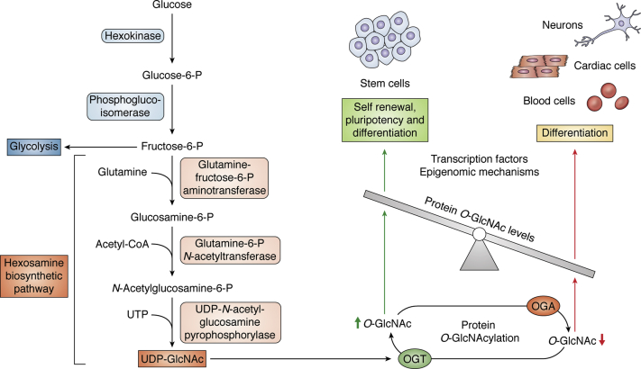
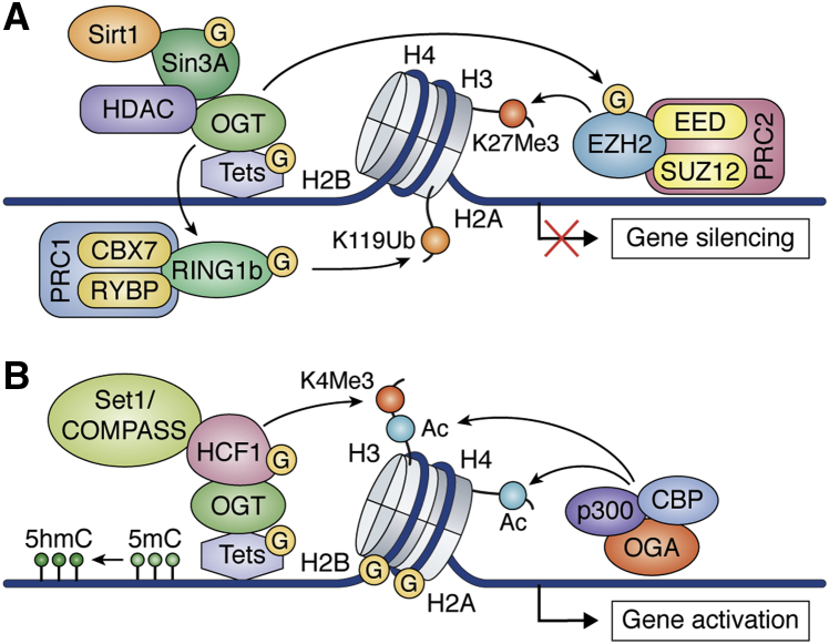
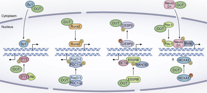
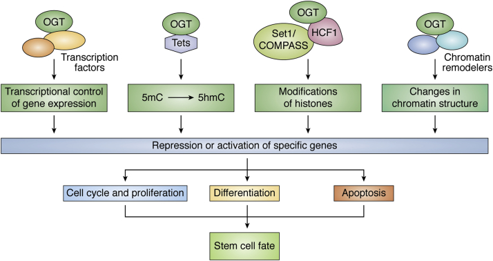
Embryonic and adult stem cells possess the capability of self-renewal and lineage-specific differentiation. The intricate balance between self-renewal and differentiation is governed by developmental signals and cell-type-specific gene regulatory mechanisms. A perturbed intra/extracellular environment during lineage specification could affect stem cell fate decisions resulting in pathology. Growing evidence demonstrates that metabolic pathways govern epigenetic regulation of gene expression during stem cell fate commitment through the utilization of metabolic intermediates or end products of metabolic pathways as substrates for enzymatic histone/DNA modifications. UDP-GlcNAc is one such metabolite that acts as a substrate for enzymatic mono-glycosylation of various nuclear, cytosolic, and mitochondrial proteins on serine/threonine amino acid residues, a process termed protein O-GlcNAcylation. The levels of GlcNAc inside the cells depend on the nutrient availability, especially glucose. Thus, this metabolic sensor could modulate gene expression through O-GlcNAc modification of histones or other proteins in response to metabolic fluctuations. Herein, we review evidence demonstrating how stem cells couple metabolic inputs to gene regulatory pathways through O-GlcNAc-mediated epigenetic/transcriptional regulatory mechanisms to govern self-renewal and lineage-specific differentiation programs. This review will serve as a primer for researchers seeking to better understand how O-GlcNAc influences stemness and may catalyze the discovery of new stem-cell-based therapeutic approaches.

Keywords: O-GlcNAcylation; cell fate determination; epigenetics; gene expression; transcription.


Conflict of interest statement

Conflict of interest The authors declare that they have no conflicts of interest with the contents of this article.

Figures

Figure 1
Figure 1
O-GlcNAcylation regulates self-renewal, pluripotency, and differentiation in stem cells. Some of glucose present inside the cells is directly channeled into HBP, where it is converted into UDP-GlcNAc, which is a key substrate for posttranslational modification of proteins through O-GlcNAcylation. The level of protein O-GlcNAcylation is determined through the dynamic activities of OGA and OGT enzymes, which in turn regulate self-renewal, pluripotency, and differentiation of stem cells via transcriptional and epigenomic control.
Figure 2
Figure 2
O-GlcNAcylation cross talk with different epigenetic modifiers to regulate gene transcription.A, OGT is targeted to promoters by TETs to recruit transcriptional repressors Sin3A, Sirt1, and HDACs to repress gene transcription. In addition, O-GlcNAcylation of RING1B by OGT at sites that are present in a region that controls the binding of RING1B to the two components of PRC1 (RYBP and CBX7) results in H2BK119 ubiquitination leading to silencing of a specific subset of genes. O-GlcNAcylated form of RINGB1 is present preferentially near genes related to neuronal differentiation of ES cells. In the PRC2 complex, EZH2 is modified by O-GlcNAcylation at Ser75, which results in it being stabilized. All of this cross talk between O-GlcNAcylation and different proteins converges onto transcriptional repression. B, O-GlcNAcylation of TET proteins results in the conversion of 5 mc to 5 hmc. OGT also modifies HCF1, which in turn recruits Set1/COMPASS to carry out H3K4me3 resulting in gene activation. At certain gene promoters, OGA recruits p300 and CBP resulting in the acetylation of H3 and H4, which results in transcriptional activation.
Figure 3
Figure 3
O-GlcNAc modifications of transcription factors affect their activities in different ways. Interaction of OGT with Sp1 results in its O-GlcNAcylation leading to nuclear localization and enhanced transcription. Runx2 is O-GlcNAcylated during osteogenic differentiation of MSCs, which results in increased activity of its transcriptional targets. The phosphorylated form of C/EBPβ is bound with DNA to carry out expression of adipogenesis-related genes. However, O-GlcNAcylation of C/EBPβ in competition with phosphorylation interferes with its function leading to reduced expression of its target genes. O-GlcNAcylation of NeuroD1 by OGT causes its translocation from the cytoplasm to the nucleus. The DNA-binding capability of Pdx-1 is enhanced following O-GlcNAcylation. High glucose is also linked to increased expression of MafA. Binding of retinoblastoma protein (Rb) with YY1 inhibits its recruitment to DNA. O-GlcNAcylation of YY1 inhibits its interaction with Rb, thus increasing the ability of YY1 to bind DNA and activate transcription. O-GlcNAc modification of coactivator PGC-1α results in the recruitment of OGT to FoxO1 promoting its O-GlcNAcylation and subsequent activation. ESSRB is modified by OGT resulting in its stabilization and enhanced transcriptional activity by interacting with OCT4 and NANOG to regulate pluripotency in mouse ESCs. HOXA1 exists in both phosphorylated and O-GlcNAcylated forms. However, the function of HOXA1 O-GlcNAcylation is not clear.
Figure 4
Figure 4
Schematic diagram showing the interaction of OGT with different epigenetic modifiers and transcription factors to regulate transcription of genes that control stem cell fate through cell cycle and proliferation, differentiation, and apoptosis.

References

    1. Conaghan J., Handyside A.H., Winston R.M., Leese H.J. Effects of pyruvate and glucose on the development of human preimplantation embryos in vitro. J. Reprod. Fertil. 1993;99:87–95. - PubMed
    1. Brinster R.L. Embryo development. J. Anim. Sci. 1974;38:1003–1012. - PubMed
    1. Shyh-Chang N., Daley G.Q., Cantley L.C. Stem cell metabolism in tissue development and aging. Development. 2013;140:2535–2547. - PMC - PubMed
    1. Heilig C., Brosius F., Siu B., Concepcion L., Mortensen R., Heilig K., Zhu M., Weldon R., Wu G., Conner D. Implications of glucose transporter protein type 1 (GLUT1)-haplodeficiency in embryonic stem cells for their survival in response to hypoxic stress. Am. J. Pathol. 2003;163:1873–1885. - PMC - PubMed
    1. Miyazawa H., Aulehla A. Revisiting the role of metabolism during development. Development. 2018;145 - PubMed

Publication types

Substances