. 2020 Nov 5;10(1):19216.
doi: 10.1038/s41598-020-76254-4.

The smart activatable P2&3TT probe allows accurate, fast, and highly sensitive detection of Staphylococcus aureus in clinical blood culture samples

Affiliations

The smart activatable P2&3TT probe allows accurate, fast, and highly sensitive detection of Staphylococcus aureus in clinical blood culture samples

Marina López-Álvarez et al. Sci Rep. .

Abstract

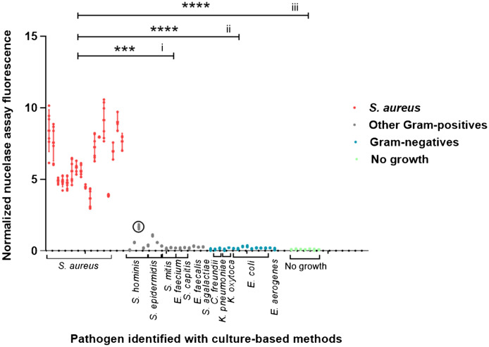
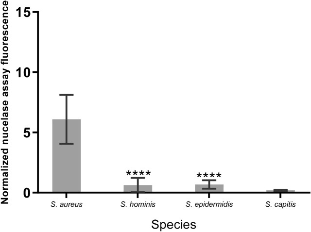
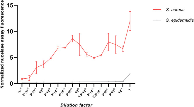
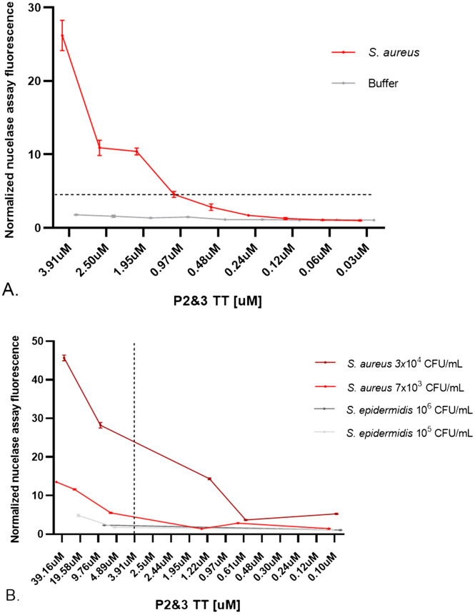
Staphylococcus aureus bacteraemia (SAB) is associated with high mortality and morbidity rates. Yet, there is currently no adequate diagnostic test for early and rapid diagnosis of SAB. Therefore, this study was aimed at exploring the potential for clinical implementation of a nuclease-activatable fluorescent probe for early diagnosis of SAB. To this end, clinical blood culture samples from patients with bloodstream infections were incubated for 1 h with the "smart" activatable P2&3TT probe, the total assay time being less than 2 h. Cleavage of this probe by the secreted S. aureus enzyme micrococcal nuclease results in emission of a readily detectable fluorescence signal. Incubation of S. aureus-positive blood culture samples with the P2&3TT probe resulted in 50-fold higher fluorescence intensity levels than incubation with culture-negative samples. Moreover, incubation of the probe with non-S. aureus-positive blood cultures yielded essentially background fluorescence intensity levels for cultures with Gram-negative bacteria, and only ~ 3.5-fold increased fluorescence intensity levels over background for cultures with non-S. aureus Gram-positive bacteria. Importantly, the measured fluorescence intensities were dose-dependent, and a positive signal was clearly detectable for S. aureus-positive blood cultures with bacterial loads as low as ~ 7,000 colony-forming units/mL. Thus, the nuclease-activatable P2&3TT probe distinguishes clinical S. aureus-positive blood cultures from non-S. aureus-positive blood cultures and culture-negative blood, accurately, rapidly and with high sensitivity. We conclude that this probe may enhance the diagnosis of SAB.


Conflict of interest statement

J.O.M. is founder and CEO of Nuclease Probe Technologies Inc., and he holds patents on oligonucleotide-based nuclease-activatable probes for detection of bacterial nucleases. G.M.v.D. is CEO, founder and shareholder of AxelaRx/TRACER group, CSO AxelaRx Biosciences Inc. The other authors have no conflicts of interest to disclose.

Figures

Figure 1
Figure 1
Evaluation of the P2&3TT probe in blood cultures. Nuclease activity assays were carried out with blood culture samples from: patients with negative-culture outcomes (n = 7, green), and patients with positive blood culture outcomes for non-S. aureus pathogens including both Gram-positive bacteria (n = 17, grey) and Gram-negative bacteria (n = 15, blue), or S. aureus (n = 17, red) (probe concentration 3.9 µM). The detected pathogens are indicated. After correction for the background based on subtraction of the fluorescence value of each blood sample without the probe, fluorescence values were normalized to the average fluorescence values of the probe signal in buffer. Variations in nuclease activity in the different blood samples are indicated by dots. Of note, a S. hominis-infected blood culture co-infected with S. saprophyticus and S. epidermidis is marked with a circle. The fluorescence intensity of this sample (1.67, SD 0.16) is higher than expected based on the T/B value for S. hominis. Possibly, this sample contained also a minor undetected fraction of S. aureus, or the detected S. hominis, S. epidermidis or S. saprophyticus secreted a nuclease that can cleave the probe. To test statistical significance, the nuclease activity measured in S. aureus-positive blood culture samples was compared with that in blood cultures testing positive for Gram-positive non-S. aureus bacteria (i), Gram-negative bacteria (ii), or negative blood culture samples (iii). **** Indicates P < 0.0001, *** indicates P = 0.0006 (Kruskal–Wallis GraphPad Prism 8.0.1).
Figure 2
Figure 2
Nuclease activity in blood cultures of Staphylococcus species. Average nuclease activity in blood cultures of S. aureus, S. hominis, S. epidermidis and S. capitis. 10 µl of blood culture supernatant was incubated with the P2&3TT probe for 1 h at 37˚C and fluorescence was measured and corrected as previously indicated. **** Indicates P < 0.0001. (Brown-Forsythe and Welch GraphPad Prism 8.0.1).
Figure 3
Figure 3
Evaluation of the P2&3TT probe in blood cultures diluted with blood from healthy volunteers. Nuclease activity assays were carried out with dilutions from S. aureus and S. epidermidis-positive blood cultures (Fig. 1) (probe concentration 3.9 µM). The initial amount of bacteria in the blood cultures (corresponding to dilution factor 1) was 7.1 × 107 CFU/mL and 2.16 × 109 CFU/mL for S. aureus and S. epidermidis respectively. After the correction for the background based on the subtraction of the fluorescence value of each blood sample without the probe, fluorescence values were normalized to the average fluorescence values of the probe signal in buffer.
Figure 4
Figure 4
Sensitivity of the P2&3TT probe. The P2&3TT probe was diluted in buffer and incubated at 37˚C for 1 h (A) in blood cultures from SAB patients, or (B) in blood cultures diluted with blood from healthy volunteers. Subsequently, fluorescence was measured. Error bars indicate standard deviations of triplicate measurements. The horizontal line represents fluorescence signal considered as positive for S. aureus based on the results presented in Fig. 1. The vertical line represents the probe concentration selected as optimal for SAB blood culture. Normalization of the fluorescence measurements was done by dividing each fluorescent measurement by the signal of each blood sample without the P2&3TT probe.

Similar articles

Cited by

References

    1. Peker N, Couto N, Sinha B, Rossen JW. Diagnosis of bloodstream infections from positive blood cultures and directly from blood samples: recent developments in molecular approaches. Clin. Microbiol. Infect. 2018;24:944–955. doi: 10.1016/j.cmi.2018.05.007. - DOI - PubMed
    1. Bearman GML, Wenzel RP. Bacteremias: a leading cause of death. Arch. Med. Res. 2005;36:646–659. doi: 10.1016/j.arcmed.2005.02.005. - DOI - PubMed
    1. Bartlett JG. Nosocomial bloodstream infections in US hospitals: analysis of 24,179 cases from a prospective nationwide surveillance study. Infect. Dis. Clin. Pract. 2004;39:309–317. doi: 10.1086/421946. - DOI - PubMed
    1. Burghardt EL, Flenker KS, Clark KC, Miguel J, Ince D, Winokur P, Ford B, McNamara JO. Rapid, culture-free detection of Staphylococcus aureus Bacteremia. PLoS ONE. 2016;11:e0157234. doi: 10.1371/journal.pone.0157234. - DOI - PMC - PubMed
    1. van Hal SJ, Jensen SO, Vaska VL, Espedido BA, Paterson DL, Gosbell IB. Predictors of mortality in Staphylococcus aureus Bacteremia. Clin. Microbiol. Rev. 2012;25:362–386. doi: 10.1128/CMR.05022-11. - DOI - PMC - PubMed

Publication types

MeSH terms