. 2020 Nov 5;10(1):19118.
doi: 10.1038/s41598-020-75933-6.

Potential involvement of Streptococcus mutans possessing collagen binding protein Cnm in infective endocarditis

Affiliations

Potential involvement of Streptococcus mutans possessing collagen binding protein Cnm in infective endocarditis

Ryota Nomura et al. Sci Rep. .

Abstract

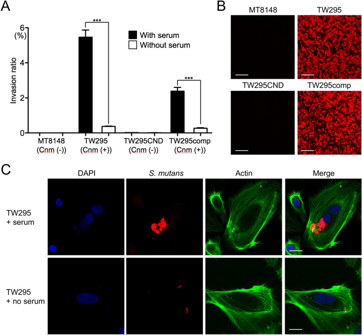
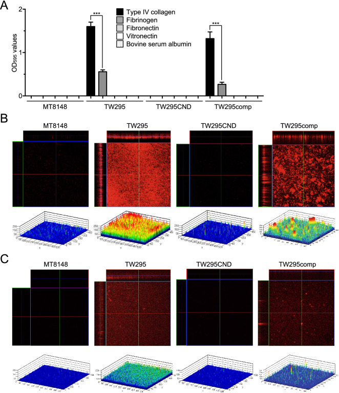
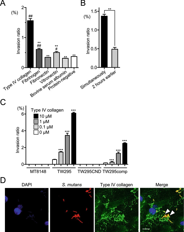
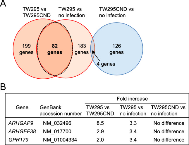
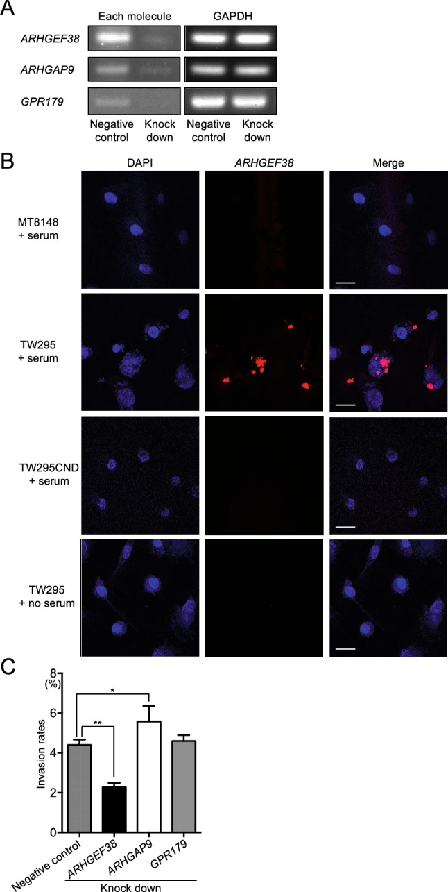
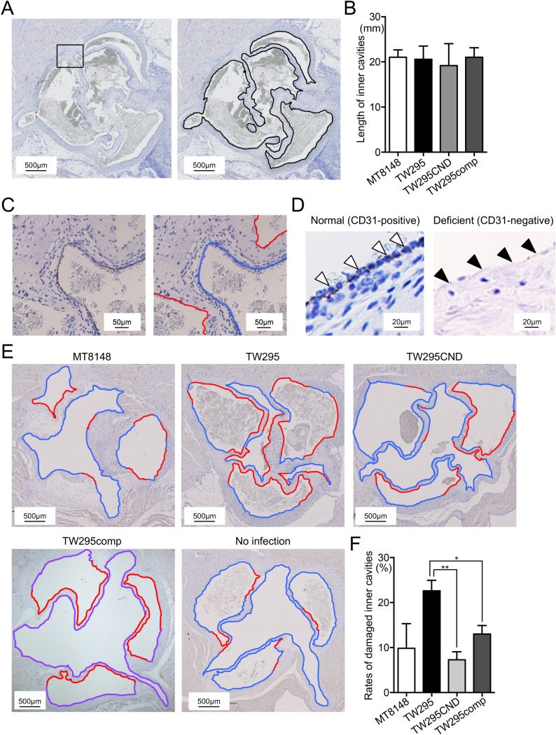
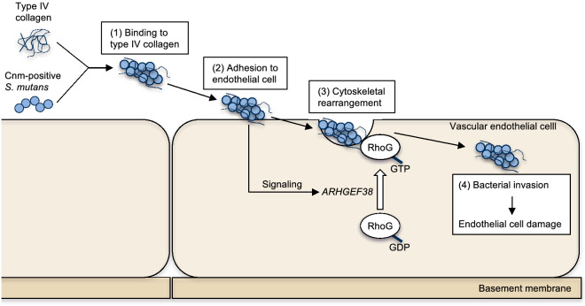
Streptococcus mutans, a significant contributor to dental caries, is occasionally isolated from the blood of patients with infective endocarditis. We previously showed that S. mutans strains expressing collagen-binding protein (Cnm) are present in the oral cavity of approximately 10-20% of humans and that they can effectively invade human umbilical vein endothelial cells (HUVECs). Here, we investigated the potential molecular mechanisms of HUVEC invasion by Cnm-positive S. mutans. The ability of Cnm-positive S. mutans to invade HUVECs was significantly increased by the presence of serum, purified type IV collagen, and fibrinogen (p < 0.001). Microarray analyses of HUVECs infected by Cnm-positive or -negative S. mutans strains identified several transcripts that were differentially upregulated during invasion, including those encoding the small G protein regulatory proteins ARHGEF38 and ARHGAP9. Upregulation of these proteins occurred during invasion only in the presence of serum. Knockdown of ARHGEF38 strongly reduced HUVEC invasion by Cnm-positive S. mutans. In a rat model of infective endocarditis, cardiac endothelial cell damage was more prominent following infection with a Cnm-positive strain compared with a Cnm-negative strain. These results suggest that the type IV collagen-Cnm-ARHGEF38 pathway may play a crucial role in the pathogenesis of infective endocarditis.


Conflict of interest statement

The authors declare no competing interests.

Figures

Figure 1
Figure 1
Invasion of HUVECs by S. mutans strains in the presence or absence of serum. (A) Invasion ratios of HUVECs after their incubation with the indicated S. mutans strains for 2 h at a multiplicity of infection of 100. Data are presented as the means ± SD of four technical replicates. ***p < 0.001 by ANOVA followed by Bonferroni’s post hoc test. (B) Representative confocal laser scanning microscopy images of S. mutans. Bacteria cells were stained red by using Alexa Fluor 555-conjugated anti-Cnm antibody. Scale bar, 10 µm. (C) Representative confocal laser scanning microscopy images of S. mutans TW295 invading HUVECs. Nuclei are stained blue (DAPI), bacteria cells are stained red (Alexa Fluor 555-conjugated anti-Cnm antibody), and actin filaments are stained green (Alexa Fluor 448-labelled phalloidin). Scale bar, 10 µm. All confocal laser scanning microscope images were taken using LSM510 (Carl Zeiss, Oberkochem, Germany).
Figure 2
Figure 2
ECM binding of S. mutans strains. (A) Binding of the indicated S. mutans strains to type IV collagen, fibrinogen, fibronectin, or vitronectin. Data are presented as the means ± SD of three technical replicates. ***p < 0.001 by ANOVA followed by Bonferroni’s post hoc test. (B, C) Confocal laser scanning microscopy images (upper panels) and schematics (lower panels) of S. mutans binding to type IV collagen (B) or fibrinogen (C). Color coding of the biofilm thickness: 0–50 µm, blue; 50–100 µm, light blue; 100–150 µm, green; 150–200 µm, yellow; and 200–250 µm, red. All confocal laser scanning microscope images and schematics were made using LSM510.
Figure 3
Figure 3
Invasion of HUVECs by S. mutans in the presence of purified ECM proteins. (A) Invasion ratios of HUVECs after their incubation with S. mutans strain TW295 for 2 h at a multiplicity of infection of 100 in the presence of type IV collagen (140 ng/ml), fibrinogen (2 mg/ml), fibronectin (0.2 mg/ml), vitronectin (500 µg/ml), or bovine serum albumin (1 mg/ml). Data are presented as the means ± SD of three technical replicates. *p < 0.05, **p < 0.01 versus TW295 and #p < 0.05, ##p < 0.01 versus the protein-negative control, using ANOVA followed by Bonferroni’s post hoc test. (B) Ability of TW295 to invade HUVECs when type IV collagen and bacteria were added simultaneously or when type IV collagen was added 2 h before the bacterial infection. Data are presented as the means ± SD of three technical replicates. **p < 0.01 using a Student’s t-test. (C) HUVEC invasion ratio (as described for A) of the indicated strains in the presence of various concentrations of type IV collagen. Data are presented as the means ± SD of four technical replicates. ***p < 0.001 versus no collagen using ANOVA followed by Bonferroni’s post hoc test. (D) Representative confocal laser scanning microscopy images of S. mutans TW295 invading HUVECs. Bacteria are stained red (Alexa Fluor 555-conjugated anti-Cnm antibody), type IV collagen is stained green (Alexa Fluor 488), and nuclei are stained blue (DAPI). Arrowheads indicate areas were type IV collagen and bacteria colocalize. Scale bar, 10 µm. All confocal laser scanning microscope images were taken using LSM510.
Figure 4
Figure 4
DNA microarray analysis of HUVEC genes involved in invasion by S. mutans. (A) Venn diagram showing the selection of genes by their differential expression (> 2.0-fold difference) in the indicated infected cells. A total of 82 genes were significantly upregulated both in TW295-infected HUVECs compared with TW295CND-infected HUVECs and in TW295-infected HUVECs compared with uninfected HUVECs but were not differentially expressed in TW295CND-infected HUVECs compared with uninfected HUVECs. (B) Three of the 82 differentially expressed genes were identified as regulators of small G proteins. Microarray data was analyzed using Agilent Feature Extraction (Agilent Technologies, Santa Clara, CA, USA).
Figure 5
Figure 5
Effect of silencing genes involved in HUVEC invasion by S. mutans. (A) RT-PCR analysis of HUVECs transfected with siRNAs targeting ARHGAP9, ARHGEF38, or GPR179. GAPDH mRNA expression was analyzed as an internal control. (B) Representative confocal laser scanning microscopy images of HUVECs invaded by S. mutans in the presence or absence of serum. Nuclei are stained blue (DAPI) and each transcript (ARHGAP9, ARHGEF38, and GPR179) is stained red (Alexa Fluor 555). Arrowheads indicate the expression of each transcript. (C) HUVECs were transfected with ARHGEF38, ARHGAP9, GPR179, or control siRNAs. Invasion ratios of these HUVECs after their incubation with S. mutans strain TW295 for 2 h at a multiplicity of infection of 100. Data are presented as the means ± SD of four technical replicates. *p < 0.05, **p < 0.01 using ANOVA followed by Bonferroni’s post hoc test. Scale bar, 10 µm. All confocal laser scanning microscope images were taken using LSM510.
Figure 6
Figure 6
Evaluation of vascular endothelial cell damage in a rat IE model. (A) Representative IHC images of anti-CD31-stained sections of extirpated heart valves from rats infected with S. mutans strain TW295. Black lines indicate the total length of the endocardium. (B) Total length of the endocardium of extirpated hearts from rats infected with the indicated S. mutans strain. Data are presented as the mean ± SD of six biological replicates per strain. (C) Magnified images of the box outlined in (A). Right panel shows endothelial cell damage. Blue and red lines indicate non-damaged (CD31-positive) and damaged (CD31-negative) endocardium, respectively. (D) Representative magnified images of normal (CD31-positive; arrowheads in left panel) and deficient (CD31-negative; arrowheads in right panel) endocardium. (E) Representative images of vascular endothelial cell damage in hearts from rats infected with the indicated S. mutans strains. Blue and red lines indicate non-damaged (CD31-positive) and damaged (CD31-negative) endocardium, respectively. (F) Rates of damaged endocardium following infection by each S. mutans strain. Data are presented as the mean ± SD of six biological replicates per strain. *p < 0.05, **p < 0.01 using ANOVA followed by Bonferroni’s post hoc test. The lengths of total, CD31-positive, and CD31-negative endocardium in the heart valves were measured using WinROOF Vol 5.0 software (Mitsuya Co., Ltd., Tokyo, Japan).
Figure 7
Figure 7
Proposed model of the interaction between Cnm-positive S. mutans and vascular endothelial cells. Cnm-positive S. mutans strains adhere to type IV collagen in the serum, and (2) then adhere to vascular endothelial cell surfaces. (3) The binding of bacteria activates ARHGEF38 in the endothelial cells, leading to GDP–GTP exchange and the activation of Rho family G proteins, cytoskeletal rearrangement, and bacterial internalization. (4) Invasion by Cnm-positive S. mutans induces vascular endothelial cell damage.

Similar articles

Cited by

References

    1. Li X, Kolltveit KM, Tronstad L, Olsen I. Systemic diseases caused by oral infection. Clin. Microbiol. Rev. 2002;13:547–558. doi: 10.1128/CMR.13.4.547. - DOI - PMC - PubMed
    1. Moreillon P, Que YA. Infective endocarditis. Lancet. 2004;363:139–149. doi: 10.1016/S0140-6736(03)15266-X. - DOI - PubMed
    1. Holland TL, et al. Infective endocarditis. Nat. Rev. Dis. Primers. 2017;2:16059. doi: 10.1038/nrdp.2016.59. - DOI - PMC - PubMed
    1. Nakatani S, et al. Japanese Circulation Society Joint Working Group. JCS 2017 Guideline on prevention and treatment of infective endocarditis. Circ. J. 2019;83:1767–1809. doi: 10.1253/circj.CJ-19-0549. - DOI - PubMed
    1. Roberts GJ, Lucas VS, Omar J. Bacterial endocarditis and orthodontics. J. R. Coll. Surg. Edinb. 2000;45:141–145. - PubMed

Publication types

MeSH terms